Angie Mueller PT, DPT is the instructor of Low Pressure Fitness and Abdominal Massage for Pelvic Floor Care, a new course on the hypopressive technique and abdominal massage for pelvic health. Join Dr. Mueller on July 27-29 in Princeton, NJ to learn more!
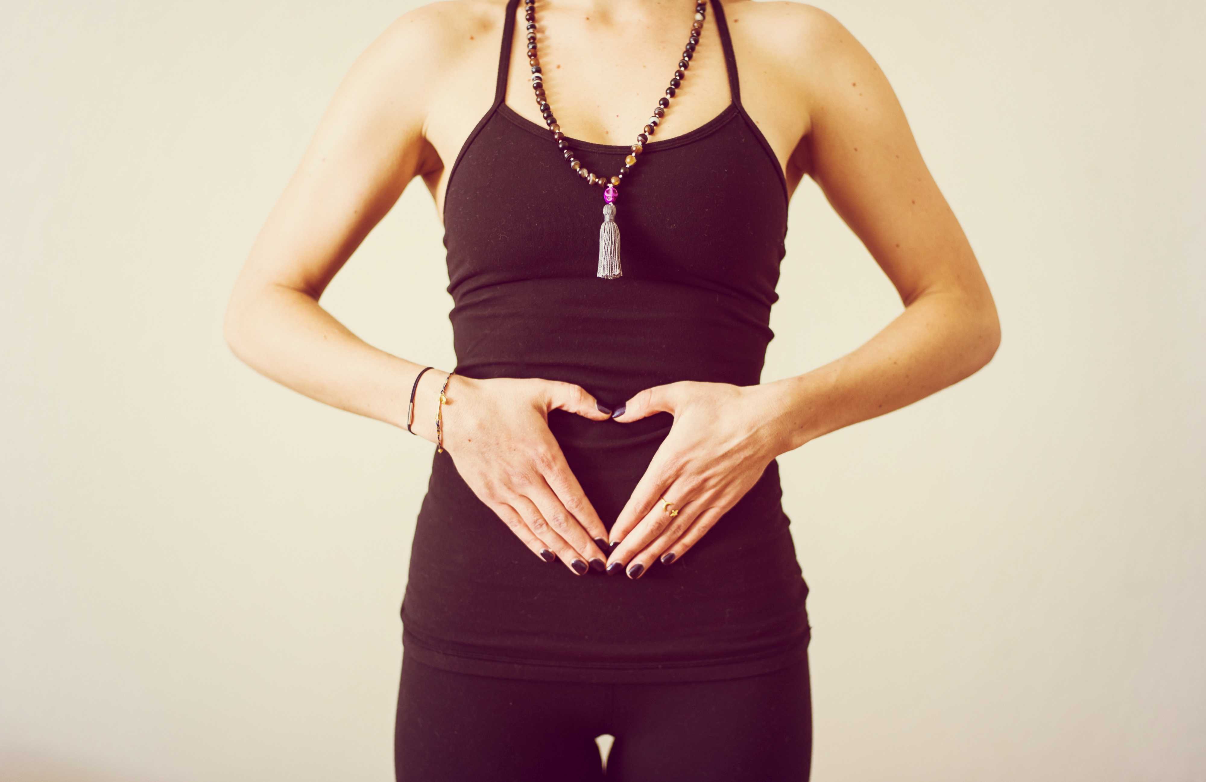
One of the first things I do as a pelvic PT when helping a woman recover from pelvic or core dysfunction, is center her uterus. I believe the uterus is the center of a women- biomechanically, physiologically, and energetically. I have seen that when the uterus is out of position, everything else in the pelvis and core is largely impacted and functions less efficiently. This includes muscular, gastrointentinal, liver, bowel and bladder, hormonal and sexual function.
 The uterus is supported by several important ligaments, which extend from the uterus out to the pelvic bones, as well as to the organs surrounding it- bladder, bowel and intestines. So if this magnificent central organ is out of her “center”- leaning forwards or backwards, or tipped to on side or the other- this can lead to a myofascial imbalance in the pelvis and cause symptoms of pelvic floor dysfunction, pain, and hormonal imbalances.
In treating thousands of women with pelvic dysfunction, I have observed that a uterus which is leaning too far forward (anteflexed) is often associated with urinary incontinence, issues with bladder urgency and frequency, and bladder prolapse (cystocele). A uterus that is tipped backwards is often associated with constipation, hemorrhoids and bowel prolapse (rectocele). A uterus that is leaning left or right is often associated with hip dysfunction, sacroiliac joint dysfunction and lumbo-pelvic alignment issues. This leads to and hip and/or knee and/or back pain due to asymmetrical pulling of the internal abdomino-pelvic fascia, especially the uterosacral and cardinal ligaments, which affects pelvic and sacral bone alignment, and then knee and ankle tracking. So centering the uterus will balance the internal pelvic and abdominal fascia, and can significantly improves cases of back pain, hip pain, knee or ankle pain.
Ensuring our organs are in their best position for receiving blood, lymph, nerve and hormonal support is critical to their health and function! If any organ in the body, especially the uterus, is not in its optimal position to receive blood, nerve, lymphatic and hormonal circulation, its function will be impacted. Therefore a mal-positioned uterus can also lead to problems with the menstrual cycle, painful periods, and fertility. When assisting any woman through a rehabilitative process, I have found it critical to appreciate how her uterine position contributes to and impacts her overall pelvic and core health- from a musculoskeletal, biomechanical and physiological perspective.
I have found that the best pelvic therapy outcomes result from use of both passive and active techniques to center the uterus. The first step is passive positioning of the uterus, which is most efficiently accomplished through abdominal massage. Abdominal self massage should be done daily. Abdominal massage will help to release any myofascial and ligamentous restrictions that are leading to a mal-positioned uterus. Abdominal massage also greatly improves blood flow and lymphatic circulation to the gut and pelvic organs leading to improved digestion and organ detoxification. Once her uterus is centered by the massage, it is important to immediately implement an active technique that will keep the uterus centered. This active uterine positioning technique must trigger the appropriate posture and breath that will keep her uterus centered with movement and throughout the activities of the day.
The second step to positioning her uterus is active activation of abdomino-pelvic musculature and key fascial chains that elevate and center the pelvic organs. This is accomplished through one of the latest core neuro-reeducation techniques- Low Pressure Fitness®. The Low Pressure Fitness methodology involves a seamless progression of postures and poses that cause a reduction in pressure in the abdomen and trigger an automatic response from the core muscles- abdominals, pelvic floor, multifidus, diaphragm. Low Pressure Fitness uses a breathing technique known as Hypopressive Breathing to reduce intra-abdominal pressure and optimize organ position. The term Hypopressive means “low pressure”. Traditional exercise, core training, sports, and most of our everyday activities are Hyperpressive – they increase the pressure in the abdomen. When the pressure in the abdomen is not appropriately managed, pressure increases, and this causes the spine to compress and the organs (especially the uterus) to move downward and away from their optimal “centered” position. But when the hypopressive breath is triggered, the pressure in the abdomen is reduced, the spine decompresses, the core musculature is gently strengthened, all of the organs lift, and the uterus is centered.
When the uterus is centered, magic happens… the fascial tension in the pelvis balances out; the resting tone of the abdominal and pelvic muscles improve and become easier to strengthen; the blood flow and lymphatic circulation in the pelvis is improved and sexual function and fertility is enhanced; hormones are better regulated and monthly cycles regulate; bowel and bladder function is optimized; the waistline reduces; pain in the back, abdomen and hips is reduced and posture improves. When all of these wonderful things occur, it is directly associated with improved energy, mood, creativity and self confidence. So remember, centering the uterus, through both active and passive techniques, is key when treating any woman. Self abdominal massage followed up by Low Pressure Fitness® are the most powerful techniques I have found to center the uterus and resolve pelvic and core dysfunction in women of all ages and lifestyles.
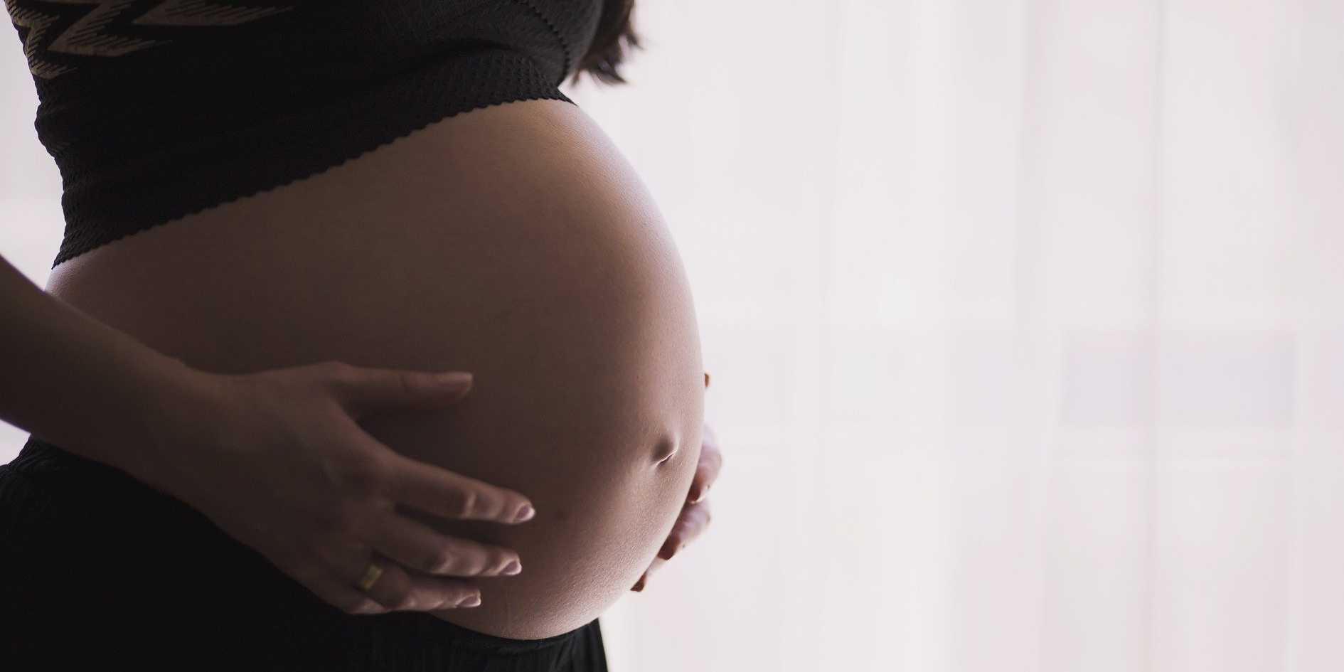
Diastasis of the Rectus Abdominis Muscle (DRAM) is the separation of the two rectus abdominis muscles along the linea alba and is very common during and after pregnancy as the rectus abdominis and linea alba stretches and thins. Patients with DRAM are often sent to seek non-surgical management for DRAM from a physical therapist (PT). Typically these patients are either at the end of their pregnancy or adjusting to round the clock care of an infant. They can be sleep deprived, and have a full schedule of doctor’s appointments, having difficulty finding childcare, making attending PT somewhat challenging. Furthermore, they may have difficulty finding the time for a home exercise program (HEP). As PT’s we often struggle with making sure to give the patient exercises that will accomplish the goal of improving DRAM, however, making sure the HEP is not so extensive or time consuming that it becomes unmanageable. Is something as easy as abdominal bracing with exercise effective for reducing DRAM in post-partum women? A recent study published in 2015 in the International Journal of Physiotherapy and Research explores just this topic(Acharry).
Why is Diastasis of the Rectus Abdominis Muscle (DRAM) important?
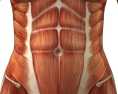
 Women with DRAM tend to have a higher degree of abdominal and pelvic region pain(Parker). Also women with DRAM may be more likely to have support related pelvic floor problem such stress urinary incontinence, fecal incontinence, or pelvic organ prolapse. The linea alba and rectus abdominis play an integral role in maintaining the anterior support of the trunk, these structures work together with pelvic girdle, posterior trunk muscles, and hips in maintaining stability when we shift weight (or transfer load) such as with standing, squatting, walking, carrying, and lifting. Therefore postural stability may be impaired with these daily tasks. Lastly, the abdominal muscles and fascia protect and support our organs so women with DRAM may have compromised support and protection of visceral structures.
Is DRAM common?
Pregnancy is the most common cause of DRAM and studies widely range from 50-100% of women experiencing DRAM at end stage pregnancy. Natural reduction and greatest recovery of DRAM usually occurs between day 1 and week 8 after delivery. Various ways exist to diagnose DRAM. The gold standard for diagnosis is computed tomography but is sometimes considered impractical due to expense. Clinically a separation of 2.0-2.7cm or “two finger widths” of horizontal separation at the umbilicus or 4.5cm above or below while performing a hooklying (supine with knees bent) abdominal curl up is considered pathological separation.
What can be done for DRAM?
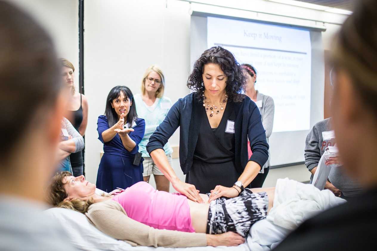
Current guidelines for conservative treatment of DRAM are sparse with little established recommendations. The earlier referenced recent cross sectional study(Acharry) explores efficacy of abdominal bracing as a treatment for reduction of DRAM in post-partum females. The study included 30 females that were one month post-partum or more who had vaginal delivery with or without episiotomy. The average distance of the diastasis was measured before and after treatment using the finger width technique. The treatment included teaching the subject four abdominal exercises, and the subjects were encouraged to complete abdominal bracing while carrying out daily activities. The four exercises included 1) static abdominal bracing exercise, lying supine with arms crossed over the diastasis for support and then pulling abdominals inwards with an isometric contraction of abdominal muscles. 2) Head lift with bracing, in hooklying with arms crossed over diastasis, exhale, lift head and use hands (or a towel/sheet) to approximate diastasis towards midline. 3) Head lift and pelvic tilt with bracing, is the same as previous exercise only adding a posterior pelvic tilt. 4) Pelvic clock exercise with bracing, visualize a clock on the lower abdominals and complete gentle movements from 12-6 o’clock, 3-9 o’clock, 12-3-6-9-12 o’clock, in a clockwise fashion and then reverse the pattern in a counter clockwise pattern. All exercises were performed twice daily, with a repetition of 5-6 days per week, for 2 weeks duration. After completing the program for two weeks the distance of the diastasis was re-measured and the average DRAM distance decreased from 3.5 to 2.5 finger widths which was considered significant. The results of this study show abdominal exercise with bracing is effective for reducing DRAM in early post partal females.
As PT’s who treat DRAM we should encourage our patients to use abdominal exercises with bracing as well as encourage our patients to use abdominal bracing with their daily tasks such as standing, lifting, weight shifting, and carrying. A home exercise program such as the one in this study proved to be effective for reducing DRAM and included only four exercises making this a manageable home exercise program for our post-partum patients.
Herman & Wallace offers a full series of peripartum courses called the "Pregnancy Series". To learn more about diastasis of the rectus abdominis muscle, join Holly Tanner, PT, DPT, MA, OCS, WCS, PRPC, LMP, BCB-PMB, CCI at Care of the Postpartum Patient in Seattle, WA this March 12-13, 2016!
Acharry, N., & Kutty, R. K. (2015). ABDOMINAL EXERCISE WITH BRACING, A THERAPEUTIC EFFICACY IN REDUCING DIASTASIS-RECTI AMONG POSTPARTAL FEMALES. Int J Physiother Res, 3(2), 999-05.
Parker, M. A., Millar, L. A., & Dugan, S. A. (2009). Diastasis Rectus Abdominis and Lumbo‐Pelvic Pain and Dysfunction‐Are They Related?. Journal of Women’s Health Physical Therapy, 33(2), 15-22.
Copyright
© Herman & Wallace, INC
All Upcoming Continuing Education Courses
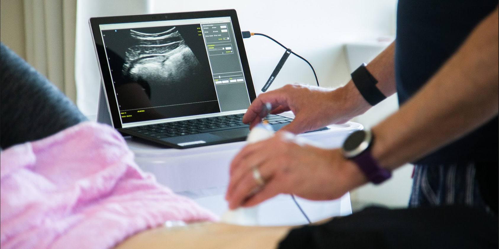
Rehabilitative Ultrasound Imaging: Pelvic Health Satellite Lab Course - Self-Hosted - April 19 - 21 2024 - SOLD OUT
Apr 19 2024 - Apr 21 2024
Rehabilitative Ultrasound: Orthopedic Topics Satellite Lab Course - Self-Hosted - April 19 - 20 2024
Apr 19 2024 - Apr 20 2024

Parkinson Disease and Pelvic Rehabilitation - Remote Course - April 19 - 20 2024
Apr 19 2024 - Apr 20 2024

Rehabilitative Ultrasound Imaging: Orthopedic Topics Satellite Lab Course - Cedar Knolls NJ - April 19 - 20 2024
Apr 19 2024 - Apr 20 2024
Rehabilitative Ultrasound Imaging: Pelvic Health Satellite Lab Course - Cedar Knolls NJ - April 19 - 21 2024
Apr 19 2024 - Apr 21 2024


Pelvic Function Level 2C - Satellite - Torrance CA - April 20 - 21 2024 - SOLD OUT
Apr 20 2024 - Apr 21 2024


Pelvic Function Level 2C - Satellite - Springfield MO - April 20 - 21 2024
Apr 20 2024 - Apr 21 2024


Pelvic Function Level 1 - Satellite - New Bern NC - April 20 - 21 2024 - SOLD OUT
Apr 20 2024 - Apr 21 2024


Pelvic Function Level 1 - Satellite - Arlington VA - April 20 - 21 2024 - SOLD OUT
Apr 20 2024 - Apr 21 2024

Pelvic Function Level 1 - Satellite - Omaha NE - April 20 - 21 2024 - SOLD OUT
Apr 20 2024 - Apr 21 2024

Pelvic Function Level 1 - Satellite - Bedford NH - April 20 - 21 2024 - SOLD OUT
Apr 20 2024 - Apr 21 2024

Pelvic Function Level 1 - Satellite - Leavenworth KS - April 20 - 21 2024 - SOLD OUT
Apr 20 2024 - Apr 21 2024

Pelvic Function Level 1 - Satellite - Boston MA - April 20 - 21 2024 - SOLD OUT
Apr 20 2024 - Apr 21 2024

Pelvic Function Level 2B - Satellite - Seattle WA - April 27 - 28 2024 SOLD OUT
Apr 27 2024 - Apr 28 2024


Pelvic Function Level 2B - Satellite - Cranford NJ - April 27 - 28 2024 - SOLD OUT
Apr 27 2024 - Apr 28 2024

Menopause Transitions and Pelvic Rehab - Remote Course - April 27 - 28 2024
Apr 27 2024 - Apr 28 2024

Nutrition Perspectives for the Pelvic Rehab Therapist - Remote Course - April 27 - 28 2024
Apr 27 2024 - Apr 28 2024


Pelvic Function Level 2B - Satellite - Des Plaines IL - April 27 - 28 2024
Apr 27 2024 - Apr 28 2024


Pelvic Function Level 2B - Satellite - Hilton Head SC - April 27 - 28 2024
Apr 27 2024 - Apr 28 2024


Pelvic Function Level 1 - Satellite - Kankakee IL - May 4 - 5 2024 - SOLD OUT
May 4 2024 - May 5 2024