Vulvar Pain: What's the Evidence?
This post was written by H&W instructor Dee Hartmann, PT, DPT. Dee will be instructing the course that they wrote on "Assessing and Treating Women with Vulvodynia" in Connecticut this September.

Within the last 4 years, two papers have been published looking at pelvic floor muscle function (PFMF) in women with and without provoked vestibulodynia (PVD). Gentilcore-Saulnier et al. sought to compare PFMF of those with and without vulvar pain and to prospectively assess how those with PVD respond PT intervention. In the first phase of their study, PFMF of 11 women diagnosed with PVD was compared to PFMF of 11 age matched controls. Outcome measures included 1) sEMG measurement of superficial and deep pelvic floor muscle (PFM) responses (both at rest and with contraction) to increasingly painful stimuli at the vulva and 2) PFMF as determined by digital internal assessment of tone, flexibility, ability to relax following contraction, and strength. In the second phase of the study, women with PVD attended 8 PT sessions (it is not clear how many were recruited from the first phase for the treatment portion and there were no controls). A specific PT protocol was utilized that included patient education, intravaginal manual therapy (including soft tissue mobilization, stretching, and desensitization), insertion activities with vaginal dilators, pelvic floor sEMG biofeedback followed by intravaginal electrical stimulation, and home exercises that included active, daily PFM exercises and every other day use of vaginal dilators.
The sEMG findings suggested that PFMF in women with PVD when compared to controls showed increased superficial PFM response to pain stimuli to the vulva but not for the deep PFMs, and significant PFM hypertonicity in the superficial but not the deep PFMs. Digital findings suggested decreased PFM flexibility and impaired ability to relax PFMs following active contraction. During intravaginal digital assessment- evaluating both superficial and deep muscles concurrently- in those with PVD, the authors found higher levels of pain with maximum voluntary contraction as well as elevated vestibular pressure sensitivity and pain intensity in both superficial and deep PFMs.
The authors concluded that women with PVD have an increased PFM response to pain and that that response was most likely due to a protective mechanism, causing active PFM contraction with impending touch, penetration, tampon insertion, fear, etc. Surface EMG showed evidence of elevated PFM activity at rest and increased PFM responsiveness in the superficial muscles following painful vulvar stimuli in those with PVD. Following 8 visits to physical therapy, women with PVD showed improvements in overall PFM tone, flexibility, strength, and ability to relax after contraction as well as improved quality of life. Patients treated also gained an increased ability to relax the PFMs following contraction which the authors hypothesize may be indicative of either increased flexibility and/or less tension at rest, or a sign of improved motor control and/or learned behavior responses gained from PT treatment.
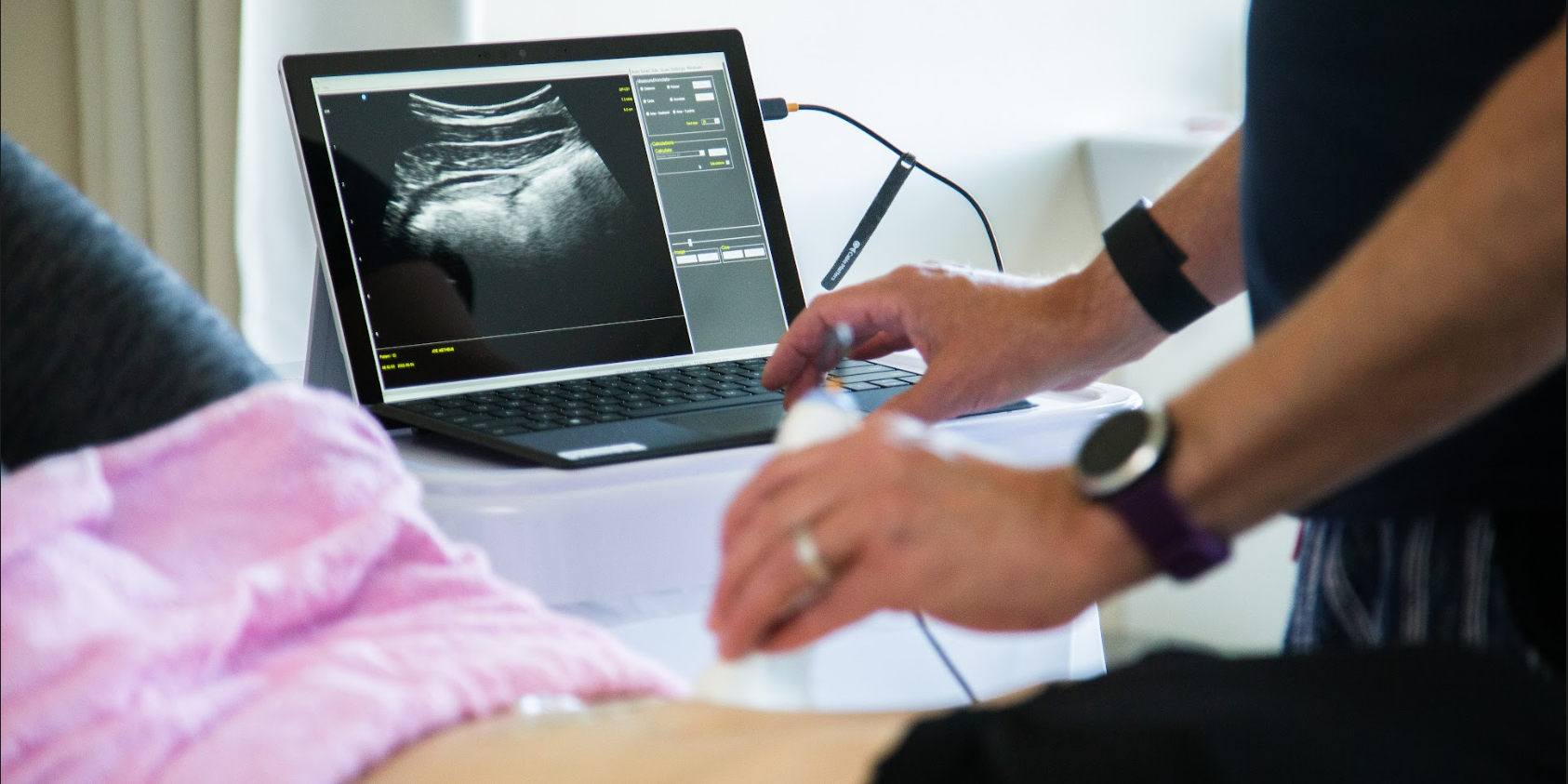
Morin et al. studied PFMF with 4D transperineal ultrasound in 49 women with PVD and 51 matched controls. Transperineal ultrasound was chosen for the ability to measure PFMF externally at the perineum without provoking pain via specific vulvar touch or internal exam (i.e. intravaginal digital or sEMG measurements.) The working hypothesis in the study was that PFM morphology at rest would differentiate those with a history of pain from controls; that is, there would be greater PFM hypertonicity in women with PVD than those without a history of pain. Ultrasound readings at rest and with maximum PFM contraction provided the main outcome measures. Parameters measured included the levator plate angle, the anorectal angle, the area of the levator hiatus, and the anterior to posterior (AP) and right to left (RL) diameters in the midsagittal and axial planes.
The resulting data in the study confirmed the hypothesis; women with PVD exhibited morphological differences from controls both at rest and with maximum contraction, suggesting elevated PFM resting tone, decreased strength, and decreased control over active PFM mobility. All parameters measured (except left to right diameter) showed statistically significant differences at rest when comparing PVD to controls as were the levator plate angle and RL diameter with contraction. When reviewing changes from baseline at rest to full contraction, all parameters measured showed statistically significant differences between the two groups—displacement of the bladder neck, levator plate and anorectal excursion, narrowing of the levator hiatus, and reduction in both the AP and RL diameters.
The conclusion of the study included that women with PVD have altered PFM morphometry at rest, suggesting an increase in PFM resting tone that appears to be chronic rather than occurring only as a defense or protective mechanism. They also suggest that those with PVD have lesser strength with maximum contraction.
Both papers provide fodder for conversation. There is very little in the literature that suggests or validates a proper treatment protocol for PFM hypertonicity. The first study cited utilized a varied treatment intervention that included daily PFM exercises, but the research lacked a control group. Authors of both papers recommended PFM retraining for women with PVD. Several retrospective reviews were published in the early 2000’s that suggested protocols including regular PFM training resulting in successful decreases in vulvar pain and improvement in sexual function and quality of life. The only published randomized, controlled trial (RCT) utilizing PFM exercise showed efficacy of all modalities when comparing vulvectomy (removal of the vulvar tissues), cognitive behavioral therapy, and sEMG education (home program included 20 minutes of PFM exercises twice daily). Report on a 10 year follow-up to the paper I published (Hartmann & Nelson, 2008) demonstrated continued improvement of symptoms, quality of life, and sexual function without further treatment. And 2/3 of the subjects were still doing PFM exercises as a home program.
What do you do with patients who have vulvar pain? Let’s talk!
Learn more about Dee Hartmann and her course by visiting Assessing and Treating Women with Vulvodynia and join her in Waterford, Connecticut next month!
References
1.Gentilcore-Saulnier E, et al. Pelvic floor muscle assessment outcomes in women with and without provoked vestibulodynia and the impact of a physical therapy program. J Sex Med. 2010;7:1003–22.
2.Morin M, et al. Morphometry of the Pelvic Floor Muscles in Women With and Without Provoked Vestibulodynia Using 4D Ultrasound. J Sex Med. 2014;11:776–785.
3.Bergeron S, et al. Physical therapy for vulvar vestibulitis syndrome: A retrospective study. J Sex Marital Ther. 2002;28:183–92.
4.Hartmann EH, Nelson CA. The perceived effectiveness of physical therapy treatment on women complaining of chronic vulvar pain and diagnosed with either vulvar vestibulitis syndrome or dysesthetic vulvodynia. Journal of the Section on Women’s Health, APTA. 25(4), Dec 2001.
5.Bergeron S, et al. A randomized comparison of group cognitive—behavioral therapy, surface electromyographic biofeedback, and vestibulectomyin the treatment of dyspareunia resulting from vulvar vestibulitis. Pain. 2001;91:297–306.
6.Hartmann D, Nelson CA. Ten years after physical therapy for vulvodynia: are there lasting benefits? J Repro Med. 2008;53(11).
By accepting you will be accessing a service provided by a third-party external to https://hermanwallace.com/
All Upcoming Continuing Education Courses
Rehabilitative Ultrasound Imaging: Pelvic Health Satellite Lab Course - Self-Hosted - April 19 - 21 2024 - SOLD OUT
Apr 19 2024 - Apr 21 2024

Rehabilitative Ultrasound Imaging: Pelvic Health Satellite Lab Course - Cedar Knolls NJ - April 19 - 21 2024
Apr 19 2024 - Apr 21 2024

Pelvic Function Level 2C - Satellite - Torrance CA - April 20 - 21 2024 - SOLD OUT
Apr 20 2024 - Apr 21 2024


Pelvic Function Level 2C - Satellite - Springfield MO - April 20 - 21 2024
Apr 20 2024 - Apr 21 2024

Pelvic Function Level 1 - Satellite - New Bern NC - April 20 - 21 2024 - SOLD OUT
Apr 20 2024 - Apr 21 2024

Pelvic Function Level 1 - Satellite - Arlington VA - April 20 - 21 2024 - SOLD OUT
Apr 20 2024 - Apr 21 2024

Pelvic Function Level 1 - Satellite - Omaha NE - April 20 - 21 2024 - SOLD OUT
Apr 20 2024 - Apr 21 2024

Pelvic Function Level 1 - Satellite - Bedford NH - April 20 - 21 2024 - SOLD OUT
Apr 20 2024 - Apr 21 2024

Pelvic Function Level 1 - Satellite - Leavenworth KS - April 20 - 21 2024 - SOLD OUT
Apr 20 2024 - Apr 21 2024

Pelvic Function Level 1 - Satellite - Boston MA - April 20 - 21 2024 - SOLD OUT
Apr 20 2024 - Apr 21 2024

Pelvic Function Level 2B - Satellite - Seattle WA - April 27 - 28 2024 SOLD OUT
Apr 27 2024 - Apr 28 2024


Pelvic Function Level 2B - Satellite - Cranford NJ - April 27 - 28 2024 - SOLD OUT
Apr 27 2024 - Apr 28 2024

Menopause Transitions and Pelvic Rehab - Remote Course - April 27 - 28 2024
Apr 27 2024 - Apr 28 2024

Nutrition Perspectives for the Pelvic Rehab Therapist - Remote Course - April 27 - 28 2024
Apr 27 2024 - Apr 28 2024


Pelvic Function Level 2B - Satellite - Des Plaines IL - April 27 - 28 2024
Apr 27 2024 - Apr 28 2024


Pelvic Function Level 2B - Satellite - Hilton Head SC - April 27 - 28 2024
Apr 27 2024 - Apr 28 2024


Pelvic Function Level 1 - Satellite - Kankakee IL - May 4 - 5 2024 - SOLD OUT
May 4 2024 - May 5 2024

Pelvic Function Level 1 - Satellite - Wichita KS - May 4 - 5 2024 - SOLD OUT
May 4 2024 - May 5 2024

Pelvic Function Level 1 - Satellite - Ann Arbor MI - May 4 - 5 2024 - SOLD OUT
May 4 2024 - May 5 2024

Pelvic Function Level 2B - Satellite - Fairfax VA - May 11 - 12 2024 SOLD OUT
May 11 2024 - May 12 2024
