
This post was written by H&W instructor Allison Ariail PT, DPT, CLT-LANA, BCB-PMD. Allison will be instructing the Pelvic Floor Level 1 course Boston this October.

Several weeks ago some of my fellow faculty members and I were discussing the resting tone of the pelvic floor. These days we take it for granted that we know there is constant low-level activity in the pelvic floor and anal sphincter in order to provide continence. However, how did this information come about? I took it upon myself to do some research to find out the beginnings of this knowledge. What I found was interesting and thought I would share.
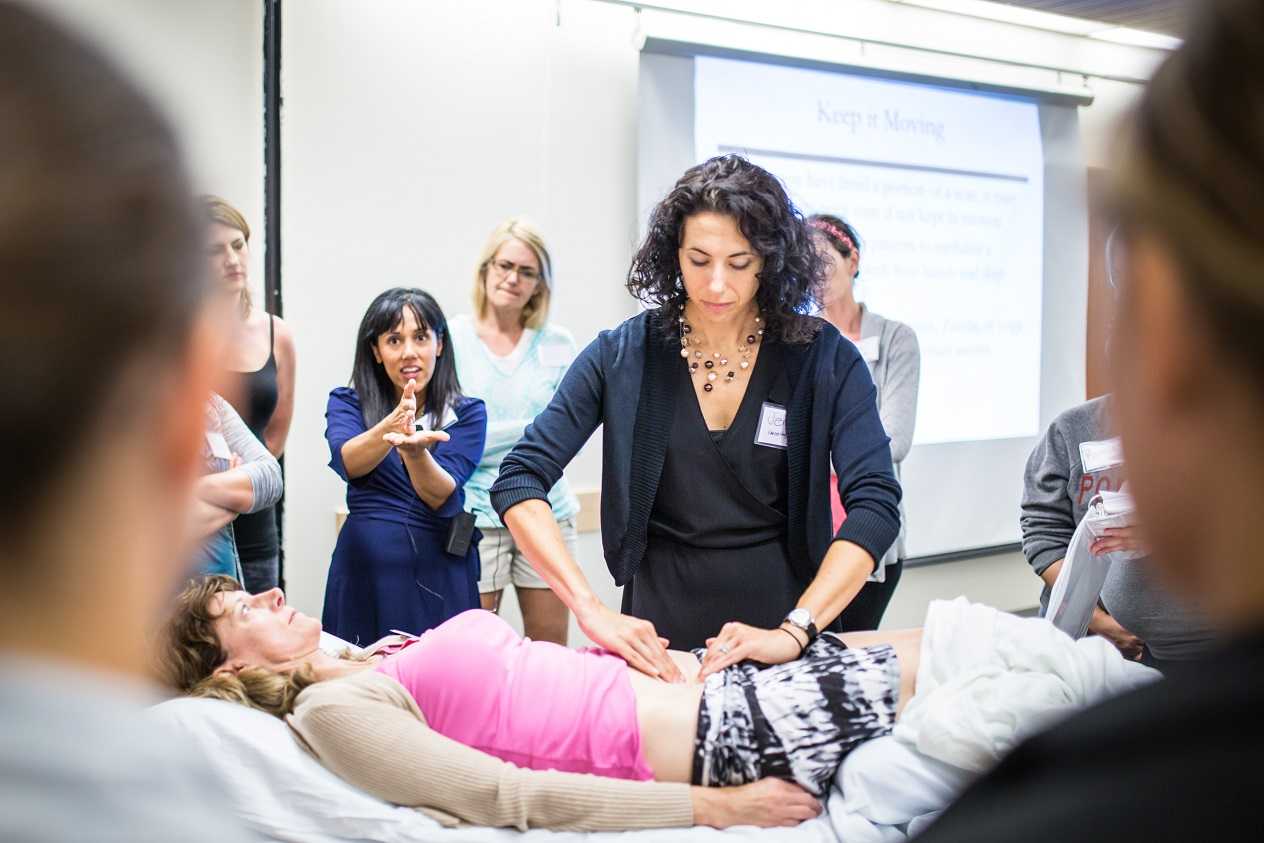
In the late 1940’s and early 1950’s the belief was held that the pelvic floor and external anal sphincters were inactive at rest, like other striated muscle throughout the body. Activity was believed to be initiated by afferent impulses from the rectal ampulla and anal canal. In 1953 Floyd and Walls found activity in the external anal sphincters at rest, even during sleep. In 1962 Parks, Porter, and Melzak published a study examining the pelvic floor muscles and the external anal sphincters using electromyography recordings. They also found activity in these muscles at rest. They hypothesized the activity was maintained by spinal reflex. These researchers looked at the activity in a healthy population, a paraplegic population, and a population that had undergone a rectal excision. When examining the paraplegic population (all subjects had complete SCI injuries above L3), they did identify activity of the pelvic floor at rest.
With respects to the rectal excision population, they examined patients whose rectums were removed, but the somatic muscles, external sphincters, and the levator ani remained with innervation intact and the muscles were sutured to provide a muscular pelvic floor. These patients also exhibited activity in the pelvic floor and anal sphincter at rest. These patients were important to the study in order to rule out that the reflex was not coming from somewhere in the rectal wall. Additionally, these researchers discovered this resting activity that was present the anal sphincter was inhibited during defecation in response to a certain degree of rectal distension.
So what did all of this new information mean to these researchers? It meant that the pelvic floor and external anal sphincter were unique due to the fact they were activated at rest, and without this activation continence would not be maintained. They determined the activation to be reflex and termed it “postural reflex of the pelvic floor.” Additionally, they termed the inhibition due to rectal distension “the rectal inhibitory reflex,” which also was due to a reflex arc. This new information was groundbreaking for the time and lead to other research that provided us with the knowledge that we have today! Thank goodness for these researchers as well as the many others who have furthered the advancement of knowledge about the pelvic floor!
Learn more about Allison and the Pelvic Floor Series by visiting our website!
1. Floyd, Walls. Electromyography of sphincter ani externus in man. J. Physiol. 122: 599, 1953.
2.Parks, Porter, Melzak. Experimental study of the reflex mechanism controlling the muscles of the pelvic floor. Dis. Colon Rectum. 5 (6): 407. 1962.
This post was written by H&W instructor Allison Ariail, PT, DPT, CLT-LANA, BCB-PMD. Allison will be instructing the Care of the Postpartum Patient course in Houston in June.
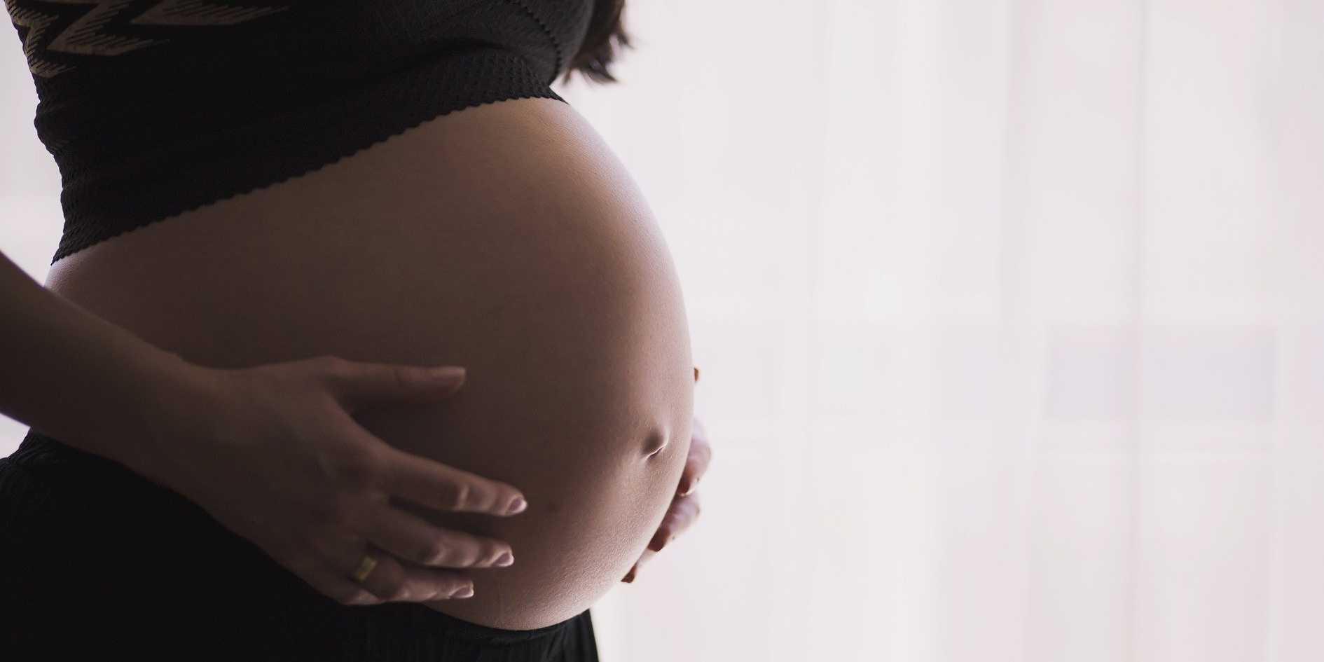
As Mother’s day weekend approaches, I take time to think about the dramatic changes in life that occur with the birth of a baby! No one is quite prepared for how much their life will change with the birth of their child, especially their first child! There are numerous changes that occur in a woman’s life during the pregnancy and into the postpartum time, both emotionally and physically. Any woman who has had a baby knows our bodies do not revert back to the exact body we had prior to pregnancy. New moms may be left with changes in their body that can greatly affect their function. Physical therapy in the postpartum time can greatly improve a woman’s well-being and function. We can treat a woman for back pain, diastasis rectus separations, incontinence, thoracic outlet syndrome, nerve damage that occurred during delivery, and many more issues a woman may present with. We also are a listening ear for the new mom going through many changes and hormonal upheaval. It is important to stay open and listen in a non-judgmental way. New moms are inundated with unsolicited advice in a way that no other patient population is. Having a safe place to come and get treated physically can help her emotionally as well.
During pregnancy and the postpartum time many habits are formed that if not changed can influence and shape how a woman lives the rest of her life. For example, night time voiding is common for pregnant women. If a woman continues to void every time she gets up with the baby in the middle of the night once she delivers, she may continue or even worsen her habit, thus creating an issue that will greatly affect her overall sleep health and well-being for the rest of her life. Having an objective person educate a woman about some of these habits can be very enlightening for an individual!
Receiving therapy in the postpartum time can influence a woman’s overall health in the immediate future as well as down the road. There are special things to consider when treating a postpartum woman and a women’s health therapist is the best person to treat her.
You can learn about special topics that affect a postpartum woman in Care of the Postpartum Patient course. The next time this course is being offered is June7-8 in Houston, Texas. So as mother’s day comes upon us, let’s celebrate the amazing journey we and other moms take in becoming a mom. Let us embrace the remarkable changes that occur physically and emotionally and thank our own mothers, as well as ourselves, for being willing to undergo these changes!

This post was written by H&W instructor Steve Dischiavi, MPT, DPT, ATC, COMT, CSCS. Dr. Dischiavi will be instructing the course that he wrote on "Biomechanical Assessment of the Hip and Pelvis" in Virginia this August.

In an outpatient sports medicine clinic the traditional model of physical therapy evaluation typically includes the therapist reviewing a patients chart and subjective symptom questionnaire of some sort. Then the therapist will bring the patient to an area to begin a subjective history and then onto a physical exam. After these procedures have been completed the therapist will typically assign a working clinical diagnosis and then begin treatment. In short, I would like to suggest a paradigm shift to this traditional model of thinking. Instead of starting the exam on a table with a static assessment of the structures involved and identifying the pain generator, I suggest the therapist begin with a specific set of movements used as an evaluative tool to identify movement dysfunction within the anatomical system as a whole.
All human interactions on earth occur between ground reaction force and gravity, our bodies are mostly just stuck in the middle of this constant battle and typically we succumb to whichever power exposes the weakest link in our biomechanical chains. One of the reasons the biomechanical chains in our bodies are so pliable and vulnerable to constant ground reaction force and gravity acting on them is because we are basically bones or struts suspended in a bag of skin all connected by soft tissue. Suggesting that without skin, fascia, and connective tissue supporting us, we would collapse to the ground in a pile of bones! Ingber (1997), suggested this concept, known as tensegrity, was the “architecture of life.” So in summary, the tensegrity structures are mechanically stable not because of the strength of the individual bones, but because of the way the entire human body distributes and balances mechanical stresses through the use of polyarticular muscle chains called slings. There will be more on slings in the upcoming blogs.
If there is truly a paradigm shift with the way we initially assess our clients and we begin our evaluations with whole system movement patterns it would be because we want to actually see how this tensgerity model is essentially collapsing under the stress of gravity. We would get a first hand glimpse in real time how energy leaks and blocks occur during human movement. These concepts are the foundation for the course Biomechanical Assessment of the Hip & Pelvis.
Ingber, D.E. (1997). Tensegrity: the architectural basis of cellular mechanotransduction.
Annual Review of Physiology, 59, 575-59.
Dr. Dischiavi's course is designed to elevate the participant’s skill level through advanced training in hip and pelvic biomechanics, functional “slings” created by the myofascial system, and through use of sports medicine theory and applied science. Biomechanical Assessment of The Hip & Pelvis will be taking place in August in Arlington, VA

There's a lot going on in the world of pelvic rehab, and continuing education is no exception! This March, Herman & Wallace is hosting NINE courses around the country. It's a lot to keep up with, so we thought you might appreciate a brief overview of what's coming up next!
Where's this pain coming from?
Pelvic pain can have many sources, and Elizabeth Hampton wants to help you quickly get to the source. Finding the Driver in Pelvic Pain empowers you to play detective in order to help even the most complex patients. Don't miss out on Finding the Driver in Pelvic Pain in San Diego, CA on March 4-6, 2016
What goes in eventually comes out
How important is a good diet? For most of us eating healthy is important, and for many pelvic rehab patients it is a necessity. That's why Megan Pribyl wrote her "Nutrition Perspectives for the Pelvic Rehab Therapist" course. This beginner level course is intended to expand the your knowledge of the metabolic underpinnings for local to systemically complex disorders. Don't miss out on Nutrition Perspectives for the Pelvic Rehab Therapist - Kansas City, MO - March 5-6, 2016!
There's fascia everywhere!
Fascial mobilization is a rising star in pelvic rehab treatment techniques, and Ramona Horton is excited to share it with you! "Mobilization of the Myofascial Layer: Pelvis and Lower Extremity" is the best opportunity you'll get to learn about the evaluation and treatment of myofascia for pelvic dysfunction. Check it out on our continuing education course page. Ramona will be teaching these techniques in Santa Barbara, CA on March 11-13.
Giving birth hurts
Sometimes the newborn is the one to get all the attention, but what about the new mother? Be sure that you can help postpartum women with symptoms like postural dysfunction, pelvic girdle dysfunction, diastasis recti abdominis and more by attending Care of the Postpartum Patient in Seattle, WA this March 12-13, taught by the wonderful Holly Tanner!
Vulvar pain is easy to have and hard to lose
12% of women in the US have vulvar pain for 3 or more months at some stage in their life. It takes a multidisciplinary approach to address all the causes and co-morbidities, and that is exactly what you'll get at Dee Hartmann's Vulvodynia: Assessment and Treatment in Houston, TX on March 12-13, 2016. Dee aims to address the vicious cycle of pain, visceral and sexual dysfunction, and the general hit to quality of life that patients with vulvodynia suffer from.
The challenge of SI joint pain
The sacroiliac joint, pelvic girdle, and pelvic ring sure can take a beating, and Peter Philip knows how to keep you moving. Through exercise and stabilization, the pelvic rehab practitioner can quickly treat pain in the lumbopelvic-hip complex. Learn all about the direct and indirect anatomy that influences the sacroiliac joint, and then get ready to find and treat the source of pain and dysfunction in Sacroiliac Joint Treatment in Minneapolis, MN on March 19-20, 2016.
Taxes and Menopause
The menopause transition is not something many people look forward to. For some women it goes more smoothly than others, and it's the less fortunate ones who need access to a well-trained pelvic care professional. Michelle Lyons is flying in from Ireland to help you to become that pro! Be it vaginal atrophy, sexual health dysfunction, pelvic organ prolapse, or any other of the myriad possible symptoms of menopause, you'll be equipped to handle them all after attending Menopause: A Rehabilitation Approach in Atlanta, GA on March 19-20, 2016.

The addition of the International Fascia Research Congress onto the scene of educational conferences has ignited an increased focus on understanding how fascia works in the body. Of course, we know fascia plays a role in compartmentalizing and separating various structures in the body, yet we also know that fascia must allow communication with the rest of the body. Is fascia simply a structural tissue that plays a mechanical role? Or does fascia hold memories, accessible during bodywork, as discussed in this article?
It may seem logical that fascia could contribute to compressive forces on the skeleton, on muscles and neurovascular structures, possibly contributing to musculoskeletal disease. Is myofascial tension sufficient to cause enough mechanical stress to create micro-damage and histochemical responses? Can this then lead to ankylosing spondylitis or axial spondyloarthritis, as discussed in this article published in Arthritis Research & Therapy? And if fascial thickness and tension is a proposed culprit of conditions such as compartment syndrome, why did these researchers find no correlation and in fact a negative correlation between fascial stiffness in patients with compartment syndrome?
Do we really know the implications of fascially-directed assessments and interventions at this time? Is the research on fascial therapy being interpreted correctly if science is still trying to figure out what fascia is, how fascia works, how fascial forces affect the body and body functions? If we don't yet understand the intricacies of the neurophysiological mechanisms that drive fascia, should we jump to conclusions about the science that may or may not be measuring the right variables? (To this end, is a test of the fascial strength meaningful if taken from a biopsy now that the tissue is disconnected from the nervous system?)
I am not a fascial researcher, and I appreciate those who do give their time and energy towards working on these questions. As a pelvic rehabilitation provider, I know that fascial relationships within the pelvis are multi-faceted and somewhat unique: the obturator internus (OI) attaches directly into a thick fascial line running between the OI and the levator ani muscles. The potential implications of this relationship on muscle strength and tension are constant clinical considerations, and ones that we hopefully will know more about as tools such as functional MRI lend improved data.
Clinicians who utilize myofascial assessments and treatment have more understanding of the role of substances such as hyaluronic acid in fascial health, yet we are still searching for accurate ways to describe how stretching and connective tissue manipulation can ease chronic pain. While we continue to explore the science behind the techniques, you can further your knowledge of fascia and fascial techniques at the continuing education course Myofascial Release for Pelvic Dysfunction, offered for the last time this year in Ohio this month.
Does wearing a pelvic belt affect the activation of the gluteus maximus and gluteus medius muscles in healthy males? Recent research asked this question, and the results, although difficult to extrapolate to other patient populations, are interesting. Surface electromyography (sEMG) amplitude was measured in 20 male patients during 6 exercises, and the amplitude during the exercise was compared to a maximum voluntary contraction. The findings demonstrated that muscle activation increased in the gluteus maximus when a pelvic belt was worn. Activation in the gluteus medius was unchanged for all exercise except during the clam exercise when the gluteus medius was noted to be more active.
Mean age in the study was 23 years, and all participants reported a lack of disease or injury. All were able to complete the exercises without pain. The 6 exercises that were instructed by an experienced physical therapist included hip clam, side lying hip abduction, single limb squat, single limb deadlift, frontal planar lunge, and frontal planar hop. Each exercise was performed 3 times, the order of exercise was randomized, and the dominant limb was used.
The authors bring up interesting points and hypotheses in relation to the sEMG findings. In a patient who presents with lumbar pain and delayed gluteus maximus activation, can a pelvic belt be utilized to improve muscle activation and therefore pelvic stability? Is adding a belt such as the COMPRESSOR belt used in this study valuable for allowing a patient to optimally complete dynamic activities, or does the belt inhibit gluteus medius activity by providing support that the muscles are supposed to provide? Most research invites us to consider the clinical implications of an intervention or a strategy, and the rehabilitation provider must assess the value of the strategy for that particular patient.
For practitioners who are interested in fine-tuning skills in lumbopelvic and hip assessment, Tracy Spitznagle, instructor in the Physical Therapy program at Washington University, will teach the Movement System Approach to Musculoskeletal Pelvic Pain: Lumbar, Hip, and SI Joint in April in Houston, TX. In this 2-day continuing education course, participants will learn to recognize movement impairment syndromes, perform movement tests, and develop a corrective exercise program based on a specific movement examination.
For some patients presenting to the pelvic rehabilitation provider, vaginal yeast infections related to Candida are an ongoing issue, a prior causative factor in pelvic muscle tension, or a potential perpetuating issue in a patient's pelvic dysfunction. A recent research article discussing candida as a chronic disease aims to propose a definition of and diagnostic criteria for women who have chronic vulvovaginal candidiasis (CVVC). This was a prospective study involving 50 women presumed to have CVVC and 42 controls. Women with CVVC were found to have the following characteristics when compared to the control group: history of a positive vaginal Candida swab, discharge, dyspareunia, soreness, swelling, cyclicity, and worsening of symptoms with antibiotics. The authors proposed that CVVC diagnosis can be made confidently utilizing 5 or more of the following: soreness, dyspareunia, positive vaginal swab (current or past prior response to antifungal medication, exacerbation with antibiotics, cyclicity, swelling, and discharge.
The authors stated reasons for wanting to categorizing and address this issue is that they had frequently observed patients with vulvovaginal candidiasis who did not present with acute or recurrent episodes, but rather with a continuous issue. The symptoms in this population tend to improve during menstruation and ease with antifungal therapy. An interesting observation made in this article is that vaginal swab test may be negative even in the presence of other symptoms. There are several proposed theories as to why patients with chronic VVC may not have positive cultures (which is required for a diagnosis of acute or recurrent VVC) including that a woman may have treated herself with anti fungal medication prior to testing, that CVVC is a hypersensitivity reaction, or that bowel Candida is what sets off the vaginal reaction. The authors also assert that a complaint of itching is not in and of itself a sensitive marker that should be used for diagnosing any type of VVC.
With issues of high cost and self-medication available over the counter (often used without proper diagnosis awareness of symptom differentiation can be useful in the pelvic rehabilitation environment. If a patient is self-medicating with topical vaginal anti fungal medication, yet presents with symptoms more consistent with chronic vulvovaginal candidiasis, the article asserts that oral medications (a daily dose for up to or more than 6 months rather than a weekly dose) is less likely to cause irritation to the involved tissues, is less expensive, and is more effective.
The diagnostic criteria used in the study for CVVC is that a patient would need to have one major and 5 minor criteria, while a presumptive diagnosis would require 1 major and 3-4 minor criteria. Major criteria includes having chronic, nonerosive, nonspecific vulvovaginitis. Minor criteria includes positive vaginal swab (present or prior soreness, cyclicity, dyspareunia, prior positive response to anti fungal therapy, worsening with antibiotics, swelling, and discharge. While medical providers are left to diagnose and prescribe the appropriate medical treatment, pelvic rehabilitation providers are able to ask appropriate questions and communicate with the patient and provider(s) about suspected symptoms and concerns. Awareness of varying causes of vaginal soreness, skin irritation, and chronic VVC adds to our level of expertise in directing patients towards efficient healing.
Chronic vulvar pain and differential diagnosis are topics covered in our Pelvic Floor Series Level 3 class. Fortunately, if you sign up quickly you may still catch one of the remaining seats in our San Diego PF3 at the end of this month!
While hysterectomy is the second most common surgery performed on women; hysterectomy rates in the US have been declining as awareness improves about minimally invasive alternatives. According to the National Women's Health Network (NWHN hysterectomy may be associated with increased risk of heart attack, surgical complications, urinary dysfunction, fistula, UTI's, sexual dysfunction, depression, and hormonal deficiencies. The NWHN describes medical necessity for hysterectomy as occurring in cases of invasive cancer, unmanageable infection or bleeding, and uterine rupture or other serious peripartum complications.
What can a woman do as an alternative to surgery? For fibroids, medication, laser ablation, cryosurgery, and myomectomy may be options available to a woman. For precancerous cells or non-cancerous growths, a LEEP procedure or cryosurgery can be performed, or a partial rather than a complete hysterectomy can be completed. Endometrial ablation or dilation and curettage (D&C) can be used to remove the lining of abnormal tissue. Endometroisis may be managed with laparoscopy, pain medication, and hormone therapy, and symptoms of a uterine prolapse may be aided by a pessary, suspension surgery, or by pelvic rehabilitation. (Hysterectomy, 2005)
In an article by Solnik and Munro (2014) indications and alternatives to hysterectomy are discussed. The authors emphasize that the physician must make every effort to determine the true etiology of the patient's pain, and they caution that women who have chronic pelvic pain "…should be counseled against hysterectomy…" In the clinical practice of the pelvic rehabilitation provider, there is value in being aware of the alternatives to the extent that we can present the current options available to a patient. Directing women to discuss alternatives to hysterectomy with their medical providers may be helpful, and directing women to websites such as the National Women's Health Network or womenshealth.gov can allow the patient to explore options for herself.
If you are interested in learning more about advanced concepts in pelvic rehabilitation such as clinical reasoning regarding patients who are candidates for hysterectomy or conservative care for symptom management, the PF3 Course in the pelvic floor series is an excellent class. Click here to find out when you can sign up for this popular course!
References
Hysterectomy. (2014). Retrieved April 16, 2014 fromhttps://nwhn.org/hysterectomy.
SOLNIK, M. J., & MUNRO, M. G. (2014). Indications and Alternatives to Hysterectomy.Clinical obstetrics and gynecology,57(1 14-42.
Prostate removal via open, laparoscopic or robotic surgical techniques has been a treatment of choice for patients with prostate cancer. Historically, patients have been keen to inquire about "nerve-sparing" procedures for prostatectomy with a goal of reducing erectile dysfunction or urinary incontinence, two common unwanted side effects of prostate surgery. Research published in Prostate International journal proposes that exquisite knowledge of fascial anatomy is a key to minimizing negative impact from surgery caused by damage to the prostatic neurovascular bundles. The authors in this paper point out that anatomical controversy exists in the literature and that the anatomy is still being investigated, increasing the surgical challenge for those physicians who aim to identify the structures.
The pelvic organs are covered by pelvic, also called endopelvic, fascia, that is commonly divided into two layers: that which covers the viscera (wrapping around each organ structure and the parietal component which covers the medial levator ani, obturator internus, and piriformis. Access to the prostate gland is gained by an anterolateral incision through the endopelvic fascia at the fusion of the visceral and parietal fascia, according to the article. Layers of prostatic fascia and the endopelvic fascia attach laterally at the tendinous arch of the pelvic fascia, and these structures attach to the puboprostatic ligaments. The puboprostatic ligaments anchor the prostate to the pubic bone, creating an important aspect of continence through fascial tension and support.
While nerve-sparing techniques have focused on preserving pelvic plexus autonomic nerve fibers, the authors argue that there is not a definite anatomy of the periprostatic nerve fibers, possibly contributing to the variability in surgical outcomes reporting for nerve-sparing procedures. Various approaches have been detailed in the literature, and are described in this article, with emphasis on dissection plane and intra- and interfascial techniques utilized.
This is a full access article with images and details beyond what most pelvic rehabilitation providers need. What is of great interest across professions is the recognized need for acute anatomical knowledge with application of skilled techniques with such anatomy in mind. The authors conclude that "…the relation of the periprostatic fascial layers on the anterior, lateral, and posterior sides of the prostate should be of great interest. A better understanding of the relation between nerve fibers and pelvic fascial layers is crucial…" Most of us were never introduced to detailed pelvic anatomy, male or female, in school. To learn more about male pelvic anatomy, you can attend either the pelvic floor series course that introduces male pelvic health, called PF2A, offered in October in St. Louis- this is the only PF2A with open seats this year. You can also attend the Male Course, offered again this year in October in Tampa.
Research led by Mei Fu, associate professor of Chronic Disease Management at New York College of Nursing, offers support for a preventive approach to lymphedema following breast cancer treatment. 140 women who were followed for 12 months were included in the study and outcomes included limb volume measurement from baseline (prior to surgery 2-4 weeks post-surgery, and at 6 and 12 months. Lymphedema was defined in the study as a 10% or greater increase in limb volume. 134 women completed the study, with 97% maintaining limb volume.
Of the subjects studied, axillary lymph node dissection was completed in almost 60%, and approximately 40% had sentinel lymph node biopsy. The self-care strategies in the research included shoulder mobility exercises, muscle-tightening deep breathing, muscle-tightening pumping exercises, and large muscle group exercise such as walking, swimming, yoga) to promote lymph health. The participants were also instructed in nutritional information aimed toward maintaining body mass index (BMI.) 97% of the women were also able to maintain BMI at the 12 month follow-up.
The majority of women in this pilot study also reported that the educational program helped in understanding of risk reduction for lymphedema, and also reduced their fear and anxiety about the condition. This type of research is very encouraging towards empowering patients following breast cancer. The authors note that a larger study population in a randomized, controlled trial will offer further information to guide clinical program development. While this study focused on participants with a diagnosis of breast cancer, is it likely that similar lifestyle and activity education would offer prevention of abdominopelvic and lower extremity lymphedema?
Within the Institute's Oncology series, you can learn more about these topics at several continuing education courses. To learn more about lymphedema, check out the Rehab for the Breast Oncology Patient, or the Oncology and the Pelvic Floor Courses (divided into male and female topics.) The next opportunity to take the Oncology for the Pelvic Floor Female: Reproductive and Gynecologic Cancers is June 21-22 in Florida. There a few seats left, so sign up soon!
All Upcoming Continuing Education Courses

Mobilization of Gastrointestinal Visceral Fascia Satellite Lab Course - Self-Hosted - May 17 - 19 2024
May 17 2024 - May 19 2024

Mobilization of Visceral Fascia: Gastrointestinal Satellite Lab Course - Torrance CA - May 17 - 19 2024
May 17 2024 - May 19 2024

Mobilization of Visceral Fascia: The Gastrointesti System Satellite Lab Course - Bedford NH - May 17 - 19 2024
May 17 2024 - May 19 2024

Mobilization of Visceral Fascia: The Gastrointestinal System - Asheville NC - May 17 - 19 2024
May 17 2024 - May 19 2024

Mobilization of Visceral Fascia: The Gastrointestinal System Satellite Lab Course - Lansing MI - May 17 - 19 2024
May 17 2024 - May 19 2024

Mobilization of Visceral Fascia: The Gastrointestinal System Satellite Lab Course - Scarborough ME - May 17 - 19 2024
May 17 2024 - May 19 2024

Mobilization of Visceral Fascia: Gastrointestinal Satellite Lab Course - Medford OR - May 17 - 19 2024
May 17 2024 - May 19 2024

Pediatrics Level 1 - Treatment of Bowel and Bladder Disorders - Remote Course - May 18 - 19 2024
May 18 2024 - May 19 2024

Pelvic Function Level 1 - Satellite - Phoenix AZ - May 18 - 19 2024 - SOLD OUT
May 18 2024 - May 19 2024

Pelvic Function Level 1 - Satellite - Anaheim CA - May 18 - 19 2024 - SOLD OUT
May 18 2024 - May 19 2024

Pelvic Function Level 1 - Satellite - Oxford AL - May 18 - 19 2024 - SOLD OUT
May 18 2024 - May 19 2024

Pelvic Function Level 1 - Satellite - McKinney TX - May 18 - 19 2024 - SOLD OUT
May 18 2024 - May 19 2024


Mobilization of Visceral Fascia: The Gastrointestinal System Satellite Lab Course - Commerce Township MI - May 17 - 19 2024
May 19 2024 - May 19 2024

Pelvic Function Level 2B - Satellite - Mission Hills CA - May 19 - 20 2024
May 19 2024 - May 20 2024


Pelvic Function Level 1 - Satellite - Frankfort IL - June 1 - 2 2024 - SOLD OUT
Jun 1 2024 - Jun 2 2024


Pelvic Function Level 1 - Satellite - Columbus OH - June 1 - 2 2024 - SOLD OUT
Jun 1 2024 - Jun 2 2024

Mobilization of the Myofascial Layer Satellite Lab Course - Self-Hosted - June 7 - 9 2024
Jun 7 2024 - Jun 9 2024

Dry Needling and Pelvic Health - In Person - 3 - Day - Lake Stevens WA - June 7 - 9 2024
Jun 7 2024 - Jun 9 2024

Mobilization of the Myofascial Layer Satellite Lab Course - Kerrville TX - June 7 - 9 2024
Jun 7 2024 - Jun 9 2024

Mobilization of the Myofascial Layer Satellite Lab Course - East Greenwich RI - June 7 - 9 2024
Jun 7 2024 - Jun 9 2024

Mobilization of the Myofascial Layer Satellite Lab Course - Conyers GA - June 7 - 9 2024
Jun 7 2024 - Jun 9 2024
Mobilization of the Myofascial Layer Satellite Lab Course - Medford OR - June 7 - 9 2024
Jun 7 2024 - Jun 9 2024

Mobilization of the Myofascial Layer Satellite Lab Course - Houston TX - June 7 - 9 2024
Jun 7 2024 - Jun 9 2024

Pelvic Function Level 1 - In-Person - New Haven CT - June 8 - 9 2024 - SOLD OUT
Jun 8 2024 - Jun 9 2024