
Recent research in The Journal of Pediatric and Adolescent Gynecology points to the alarming number of young women who present with pelvic pain who in fact also have endometriosis. Dr. Opoku-Anane and Dr. Laufer report that prevalence rates of endometriosis in an adolescent gynecology population have likely been underestimated (reported range of 25-47%) and that with advanced surgical methods the rates have been estimated to be as high as 73% in those who have pelvic pain. In their retrospective study, 117 subjects ages 12-21 completed laparoscopic examination for endometriosis. These subjects did not previously respond to non-steroidal anti-inflammatories or to oral contraceptives, and they were all referred for evaluation of chronic pelvic pain. In addition to collecting data about patient symptoms, the stage and descriptions of any endometrial lesions were documented.
A remarkable 115 of the 117 subjects (98%) presented with Stage I or II endometriosis as defined by the American Society for Reproductive Medicine guidelines. (Click here for the link to a detailed patient education document from the ASRM that describes endometriosis as well as staging.) The median age for onset of menarche in this population was 12 years old, and the median age of first symptoms reported occurred at age 13. Nearly 16% of the subjects also reported gastrointestinal complaints, menstrual irregularity in nearly 8%, and 76% of the participants reported a family history that included endometriosis, severe dysmenorrhea, and/or infertility. The authors of this research point out that advances made in surgical technique, both from a technological standpoint and a physician skill level, may be contributing factors in the increased rates of diagnosis of endometriosis.The authors also point out that it is yet unknown if early diagnosis and treatment will lead to improved outcomes in this population.
If you are interested in learning more about endometriosis in general, click here to follow the link to a free, full text article in PubMed Central. The article was first published in 2008, and even though advances in surgical diagnosis have been made, most of the information related to symptoms, medical treatment, and related risks remain significantly unchanged. In relation to etiology of endometriosis, one study that has set forth an environmental risk for endometriosis can be accessed here. Dr. C. Matthew Peterson, one of the researchers involved with the ENDO study, presented at the 2011 International Pelvic Pain Society meeting, and he encouraged all present to consider implementing strategies to minimize risks from chemicals in our daily lives. The Environmental Protection Agency offers advice towards protecting our health that can be accessed here. If environmental hazards are influencing the onset or progression of conditions such as endometriosis, it is in our best interest to reduce these risks. Consider not only the product exposure at home, but also at the workplace, and request less toxic products including cleaners when able.
In relation to pelvic rehabilitation, patients who present with pelvic pain or other pelvic health issues due to endometriosis often find relief when working with pelvic rehab providers. While surgery may be critical in reducing severe adhesions, maximizing tissue health and patient mobility and function is a job in which we can all actively participate. The evaluation and treatment of pelvic pain is instructed at various levels of depth in all of the main series courses as well as in many other courses offered at the Herman & Wallace Pelvic Rehabilitation Institute.

This post was written by H&W instructor Michelle Lyons, PT, MISCP, who authored and instructs the course, Special Topics in Women’s Health: Endometriosis, Infertility & Hysterectomy. She will be presenting this course this February!

Endometriosis is a common gynaecological disorder, affecting up to 15% of women of reproductive age. Because endometriosis can only be diagnosed surgically, and also because some women with the disease experience relatively minor discomfort or symptoms, there is some controversy regarding the estimates of prevalence, with some authorities stating that as many as one and three women may have endometriosis (Eskenazi & Warner 1997)
There is a wide spectrum of symptoms of endometriosis, with little or no correlation between the acuteness of the disease and the severity of the symptoms (Oliver & Overton 2014). The most commonly reported symptoms are severe dysmenorrhoea and pelvic pain between periods. Dyspareunia, dyschezia and dysuria are also commonly seen. These pain symptoms can be severe and have been reported to lead to work absences by 82% of women, with an estimated cost in Europe of €30 billion per year (EST 2005). Secondary musculoskeletal impairments caused by may include: lumbar, sacroiliac, abdominal and pelvic floor pain, muscle spasms/ myofascial trigger points, connective tissue dysfunction, urinary urgency, scar tissue adhesion and sexual dysfunction (Troyer 2007) – all of which may be responsive to skilled pelvic rehab intervention.
Endometriosis can lead to inflammation, scar tissue and adhesion formation and myofascial dysfunction throughout the abdominal and pelvic regions. This can set up a painful cycle in the pelvic floor muscles secondary to the decrease in pelvic and abdominal organ/muscle/fascia mobility which can subsequently lead to decreased circulation, tight muscles, myofascial trigger points, connective tissue dysfunction and pain and possible neural irritation.
Abdominal trigger points and pain can be commonly seen after laparascopic surgery for diagnosis or treatment. We know that fascially, the abdominal muscles are closely connected with the pelvic floor muscles and dysfunction in one group may trigger dysfunction in the other, as well as causing associated stability, postural and dynamic stability issues.
The pain created by muscle tension and dysfunction, may lead to further pain and increasing central sensitisation and further disability. Unfortunately for the endometriosis patient, as well as dealing with the problems already associated with endometriosis, she may also develop a spectrum of secondary musculo-skeletal problems, including pelvic floor dysfunction – and for some patients this may actually be responsible for the majority of their pain (Troyer 2007).
The skilled pelvic rehab therapist has much to offer this under-served patient population in terms of reducing pain and dysfunction, educating regarding self-care and exercise and helping to restore quality of life. Interested in learning more? Join me for my new course: ‘Special Topics in Women’s Health: Endometriosis, Infertility & Hysterectomy’ in San Diego this February or Chicago in June.
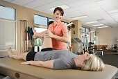
After pushing a double stroller for a 3 mile run to the park yesterday, I had a flare up of hip pain that made me doubt my ability to get my kids back home. While they were playing, a hanging ladder caught my eye and sent my manual therapy wheels spinning. I carefully slipped my leg over one of the rungs, angled my body just right, and leaned away to distract my hip. I noticed a toddler staring at me, so I politely told her, “I’m just mobilizing my hip joint, sweetie, but you can go ahead and climb now!” My relief was almost immediate, and I realized my patients need to know how to help themselves, too! We all know how to prescribe home exercises for patients regarding stretching and strengthening, but once a therapist is competent performing joint mobilizations, the need for this arthokinematic movement is often found to be essential prior to the osteokinematic movement of stretching. The hip joint in particular is affected by pathologies of the lumbar spine, the sacroiliac joint, and the pelvic floor. When the hip joint is not moving around the proper physiological axis, then the knee can be negatively impacted as well as the areas just mentioned.
 Therapists need to discern whether a patient is appropriate for self-hip mobilization instruction, as a “motor moron” probably would not be a good candidate to whom you would explain how to perform mobilizations at home. When you realize a patient “gets it,” then you can suggest the techniques to that patient. Reiman and Matheson (2013) presented a paper regarding suggestions for self-mobilization of the hip joint. They demonstrate an inferior-posterior hip glide with a towel, weight, and a step with or without muscle reeducation in hip flexion; an inferior and lateral glide with hip flexion movement; a hip posterior glide with or without movement; a hip lateral glide with or without muscle reeducation; a hip anterior glide with or without muscle reeducation; and, a long axis distraction mobilization. The authors conclude the efficacy of their protocol and techniques are not completely backed up by evidence yet and recommend they be implemented as an adjunct to evidence based practice, not a primary treatment approach.
Regarding the efficacy of hip mobilization in the clinic by a skilled clinician, a study by Makofsky et al, (2007) discusses the effect of inferior hip joint mobilization on hip abductor force. This study leaves little doubt that mobilizing the hip can facilitate contraction of the gluteus medius. A 17.35% increase in hip abduction torque was noted immediately after the inferior Grade IV hip mobilization; whereas, the control group without mobilization experienced a 3.68% decrease in hip abduction torque. We generally see patients much less often than our services are needed, so being able to teach patients how to mobilize on their own to supplement our work could be extremely effective in the long run.
I have the extreme fortune of being married to a manual therapist, so I do not always have to find crafty ways to mobilize my own joints, but my recent experience was encouraging to know it is more than possible to help myself. My hip pain had caused some patellofemoral symptoms because my gluteal muscles were inhibited. Performing a self-distraction close to my hip joint helped kick in the muscles required for greater stability of my knee. I cruised home with the kids without a hitch. We should all be ready to educate our patients to take potentially embarrassing measures to help themselves as well.
Reiman, M. P., & Matheson, J. W. (2013). RESTRICTED HIP MOBILITY: CLINICAL SUGGESTIONS FOR SELF‐MOBILIZATION AND MUSCLE RE‐EDUCATION. International Journal of Sports Physical Therapy, 8(5), 729–740.
Makofsky, H., Panicker, S., Abbruzzese, J., Aridas, C., Camp, M., Drakes, J., … Sileo, R. (2007). Immediate Effect of Grade IV Inferior Hip Joint Mobilization on Hip Abductor Torque: A Pilot Study. The Journal of Manual & Manipulative Therapy, 15(2), 103–110.

Inflammatory bowel diseases such as ulcerative colitis and Crohn’s disease (CD), according to Lan et al., are characterized by chronic inflammation in intestinal mucosa. There is little information available based on human studies that links nutritional support with inflammatory bowel disease. The authors of this article analyzed the available information about supplements that appear beneficial in healing the gut mucosa.
 The intestinal epithelium acts as a selective barrier, and inflammatory bowel disease can disrupt this important barrier, affecting absorption, mucus production, and enteroendocrine secretion. Intestinal wound healing, according to the linked article, is “dependent on the precise balance of several processes including migration, proliferation, differentiation, and apoptosis of the epithelial cells.” Although there are few human clinical studies (more are animal and cell model studies) there is evidence that both macronutrient and micronutrient deficiencies may exist in patients who have inflammatory bowel disease. Patients may have malnutrition of proteins, of minerals and vitamins. Following are some points from the article:
- Arginine, glutamine, and glutamate are noted as amino acids that contribute to mucosal healing in animal studies, as well as threonine, methionine, proline, serine, and cysteine.
- Short-chain fatty acids, which are the result of metabolized dietary fiber (grains, legumes, fruits and vegetables), and resistant starch (beans, potatoes, brown rice) are a major source of energy for epithelial cells in the colon
- Vitamin A deficiency is common in patients with newly diagnosed IBD
- Vitamin D may decrease intestinal fibrosis, and reduce risk of Crohn’s relapse
- Vitamin C and Zinc may both help heal epithelial wounds in the colon
- During the remission phase, some foods may be poorly tolerated, such as dairy
- During an active flare of IBD, a patient may require enteral nutrition
The authors conclude that the exact mechanisms by which the various dietary compounds contribute to bowel healing is still unknown. Furthermore, the ability to make clear dietary recommendations is limited by the lack of clinical studies. At a minimum, this information can alert the pelvic rehab provider to discuss the potential benefits of nutrition on bowel health with patients. Many of us are quite comfortable giving advice about drinking appropriate amounts and types of fluid, or eating more whole and less processed foods. We can take our nutritional education a step further by encouraging a patient to discuss healing supplements with a provider and/or nutritionist to best support the healing bowel. If you are interested in learning more about nutrition and pelvic health, you might love our newer course Nutrition Perspectives for the Pelvic Rehab Therapist Kansas City in March of next year with instructor Megan Pribyl.
Lan, A., Blachier, F., Benamouzig, R., Beaumont, M., Barrat, C., Coelho, D., & Tomé, D. (2015). "Mucosal healing in inflammatory bowel diseases: is there a place for nutritional supplementation?" Inflammatory bowel diseases, 21(1), 198-207.
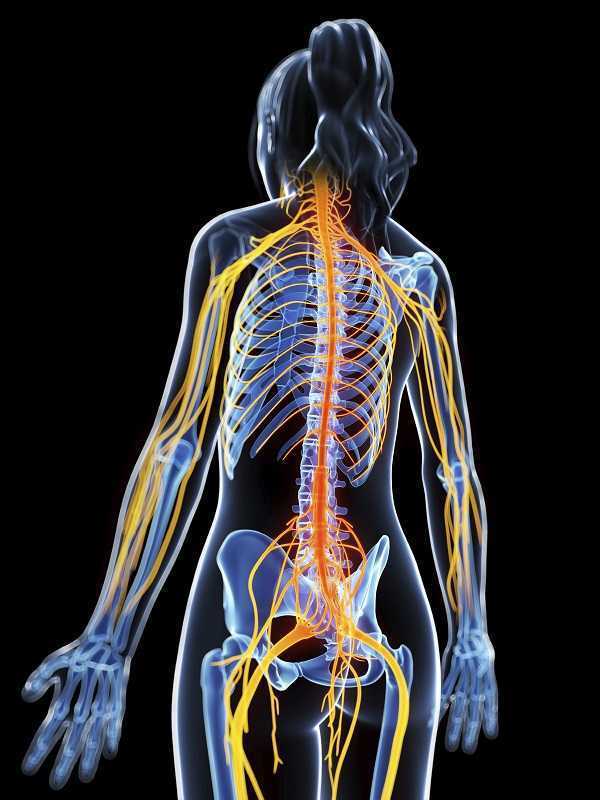
Pudendal nerve dysfunction, when severe, is truly one of the most difficult conditions treated by pelvic rehabilitation providers. While peripheral nerve dysfunction anywhere in the body can be challenging to treat, access to the nerve along its many potential sites of irritation is limited when compared to other peripheral nerves. Many research studies have been completed that investigate how structures like the median nerve move in the body, and to what extent the nerve movement changes in cases of dysfunction, yet we still have very little to work with regarding the pudendal nerve. Little, that is, except anatomical knowledge, nerve and tissue mapping and palpation skills, expert listening and evaluation skills, and an abundance of existing and emerging methodology directed to treatment of chronic pain conditions.
The Neuro Orthopaedic Institute (also known as the NOI group)has led the physiotherapy world in seeking and sharing knowledge about the evaluation and treatment of conditions involving the nervous system. In a prior posting within the "noinotes" available as a newsletter from the NOI group, the following is stated: "…for the best clinical exposure of a peripheral nerve problem, take up the part that you think holds the problem first and then progressively add tension to the nerve via the limbs." Let's say, for example, that you gently tension the pudendal nerve by completing an inferior compression of the right levator ani muscle group (towards the lateral portion of the muscle belly versus at the midline). At this point, what limb movement should be performed to increase tension to the nerve? Does a straight leg raise tension the nerve, or hip rotation, hip adduction? What evidence do we have that this nerve tension increases in terms of elongation of the peripheral nerve, and by what connective tissue attachments is this tension proposed to occur? And for using order of movement in the clinic, do we start with a pelvic muscle bearing down or contraction, then add trunk or limb movements?
The "Ordering nerves" post describes listening "…to the patient about the sequence of movements which aggravate them.." so that with clinical reasoning, for evaluation or treatment, the nerve symptoms can be reproduced to an appropriate extent. For example, if a pelvic muscle contraction significantly aggravates a patient's nerve-like symptoms, why should a patient be instructed, or allowed even, to do Kegel muscle exercises to a degree that causes significant pain? If a patient has low grade, annoying symptoms that are only reproduced with posterior pelvic floor stretch combined with an anterior pelvic tilt and passive straight leg raise with internal rotation of the hip, then that position should be incorporated into a clinical and a home program if able.
Just because we don't yet know how patients with true pudendal nerve dysfunction present clinically in terms of nerve gliding ability, and what movements typically engage particular portions of the nerve (such as the proximal portion in the posterior pelvis, the portion that lives along the obturator internus, the portion housed by the Alcock's canal, or even the longest portion of the nerve that extends to the genitals), that does not mean we should default to a one-size-fits-all pelvic muscle strengthening or stretching approach. Each patient must be met with curiosity, and with keen knowledge of anatomy, nerve evaluation principles, and pain-brain centered skills so that an individual approach is designed. As is concluded in this post from the NOI group, we must "Keep playing with order of movement."
If you would love to fill up your toolbox with concepts and techniques for treating pudendal nerve dysfunction, sign up quickly for the last chance this year to take Pudendal Neuralgia and Treatment in San Diego this August.
Postpartum perineal injuries can cause pain and dysfunction for a short or an extended period of time. Pelvic rehab providers are in a position to educate women about the immediate and long-term management of perineal pain. A 2015 study by Manfre et al. assessed the response of cortisone cream application to the perineum in the immediate postpartum period. The study was a randomized controlled trial involving 27 subjects with each subject serving as her own control. Three different treatments were given over a 12 hour period: corticosteroid, placebo, and no treatment. (The hydrocortisone cream was at 1% in an alcohol-based cream, the placebo was a non-medicated acetyl alcohol-based cream.) The cream was applied by an investigator who placed the cream on a Witch Hazel pad. The participants and the researchers were blinded to the type of cream applied, and the applications were randomized and took place within the first 12.5 hours after birth. Perineal pain levels were assessed immediately before cream application, and at 30 and 60 minutes after application. Using a visual analog scale (VAS), the symptom of pain was assessed and compared to baseline. In the study, the authors report that in the immediate postpartum period, women in their institution were often prescribed medication ranging from ibuprofen to hydrocodone. Topical medications, ice packs, heat packs are also mentioned as available treatments. Other pain medications or cold packs were available during the study; no other topical creams were utilized.
 Results indicated that the participants responded positively to both creams with significantly more pain reduction than the no treatment group. The authors propose that both creams provided a soothing effect by providing moisture to the tissues, creating a protective barrier, and preventing friction and irritation. Because the placebo emollient cream was not significantly more expensive than the hydrocortisone cream, the article suggests using hydrocortisone on the postpartum perineum due to the medication’s potential beneficial anti-inflammatory effects. Also of note was that ice packs were used by less than half of the women in the study, and when ice was used, it was only during the first four hours after birth. One reason for the low frequency use of ice was thought to be the difficulty in maintaining ice application on the perineum.Manfre 2015
Because the pelvic rehab provider is in an optimal role as educator for pain reduction strategies, this study provides some interesting information to share with other birth providers and with patients. Learn more about postpartum patient care at Care of the Postpartum Patient, available in Seattle this March.
Manfre, M., Adams, D., Callahan, G., Gould, P., Lang, S., McCubbins, H., ... & Chulay, M. (2015). Hydrocortisone Cream to Reduce Perineal Pain after Vaginal Birth: A Randomized Controlled Trial. MCN: The American Journal of Maternal/Child Nursing, 40(5), 306-312.
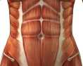
Diastasis of the Rectus Abdominis Muscle (DRAM) is the separation of the two rectus abdominis muscles along the linea alba and is very common during and after pregnancy as the rectus abdominis and linea alba stretches and thins. Patients with DRAM are often sent to seek non-surgical management for DRAM from a physical therapist (PT). Typically these patients are either at the end of their pregnancy or adjusting to round the clock care of an infant. They can be sleep deprived, and have a full schedule of doctor’s appointments, having difficulty finding childcare, making attending PT somewhat challenging. Furthermore, they may have difficulty finding the time for a home exercise program (HEP). As PT’s we often struggle with making sure to give the patient exercises that will accomplish the goal of improving DRAM, however, making sure the HEP is not so extensive or time consuming that it becomes unmanageable. Is something as easy as abdominal bracing with exercise effective for reducing DRAM in post-partum women? A recent study published in 2015 in the International Journal of Physiotherapy and Research explores just this topic(Acharry).
Why is Diastasis of the Rectus Abdominis Muscle (DRAM) important?
 Women with DRAM tend to have a higher degree of abdominal and pelvic region pain(Parker). Also women with DRAM may be more likely to have support related pelvic floor problem such stress urinary incontinence, fecal incontinence, or pelvic organ prolapse. The linea alba and rectus abdominis play an integral role in maintaining the anterior support of the trunk, these structures work together with pelvic girdle, posterior trunk muscles, and hips in maintaining stability when we shift weight (or transfer load) such as with standing, squatting, walking, carrying, and lifting. Therefore postural stability may be impaired with these daily tasks. Lastly, the abdominal muscles and fascia protect and support our organs so women with DRAM may have compromised support and protection of visceral structures.
Is DRAM common?
Pregnancy is the most common cause of DRAM and studies widely range from 50-100% of women experiencing DRAM at end stage pregnancy. Natural reduction and greatest recovery of DRAM usually occurs between day 1 and week 8 after delivery. Various ways exist to diagnose DRAM. The gold standard for diagnosis is computed tomography but is sometimes considered impractical due to expense. Clinically a separation of 2.0-2.7cm or “two finger widths” of horizontal separation at the umbilicus or 4.5cm above or below while performing a hooklying (supine with knees bent) abdominal curl up is considered pathological separation.
What can be done for DRAM?
Current guidelines for conservative treatment of DRAM are sparse with little established recommendations. The earlier referenced recent cross sectional study(Acharry) explores efficacy of abdominal bracing as a treatment for reduction of DRAM in post-partum females. The study included 30 females that were one month post-partum or more who had vaginal delivery with or without episiotomy. The average distance of the diastasis was measured before and after treatment using the finger width technique. The treatment included teaching the subject four abdominal exercises, and the subjects were encouraged to complete abdominal bracing while carrying out daily activities. The four exercises included 1) static abdominal bracing exercise, lying supine with arms crossed over the diastasis for support and then pulling abdominals inwards with an isometric contraction of abdominal muscles. 2) Head lift with bracing, in hooklying with arms crossed over diastasis, exhale, lift head and use hands (or a towel/sheet) to approximate diastasis towards midline. 3) Head lift and pelvic tilt with bracing, is the same as previous exercise only adding a posterior pelvic tilt. 4) Pelvic clock exercise with bracing, visualize a clock on the lower abdominals and complete gentle movements from 12-6 o’clock, 3-9 o’clock, 12-3-6-9-12 o’clock, in a clockwise fashion and then reverse the pattern in a counter clockwise pattern. All exercises were performed twice daily, with a repetition of 5-6 days per week, for 2 weeks duration. After completing the program for two weeks the distance of the diastasis was re-measured and the average DRAM distance decreased from 3.5 to 2.5 finger widths which was considered significant. The results of this study show abdominal exercise with bracing is effective for reducing DRAM in early post partal females.
As PT’s who treat DRAM we should encourage our patients to use abdominal exercises with bracing as well as encourage our patients to use abdominal bracing with their daily tasks such as standing, lifting, weight shifting, and carrying. A home exercise program such as the one in this study proved to be effective for reducing DRAM and included only four exercises making this a manageable home exercise program for our post-partum patients.
Herman & Wallace offers a full series of peripartum courses called the "Pregnancy Series". To learn more about diastasis of the rectus abdominis muscle, join Holly Tanner, PT, DPT, MA, OCS, WCS, PRPC, LMP, BCB-PMB, CCI at Care of the Postpartum Patient in Seattle, WA this March 12-13, 2016!
Acharry, N., & Kutty, R. K. (2015). ABDOMINAL EXERCISE WITH BRACING, A THERAPEUTIC EFFICACY IN REDUCING DIASTASIS-RECTI AMONG POSTPARTAL FEMALES. Int J Physiother Res, 3(2), 999-05.
Parker, M. A., Millar, L. A., & Dugan, S. A. (2009). Diastasis Rectus Abdominis and Lumbo‐Pelvic Pain and Dysfunction‐Are They Related?. Journal of Women’s Health Physical Therapy, 33(2), 15-22.
Copyright
© Herman & Wallace, INC
 Sexual dysfunction is a common negative consequence of Multiple Sclerosis, and may be influenced by neurologic and physical changes, or by psychological changes associated with the disease progression. Because pelvic floor muscle health can contribute to sexual health, the relationship between the two has been the subject of research studies for patients with and without neurologic disease. Researchers in Brazil assessed the effects of treating sexual dysfunction with pelvic floor muscle training with or without electrical stimulation in women diagnosed with multiple sclerosis (MS.) Thirty women were allocated randomly into 3 treatment groups; 20 women completed the study. All participants were evaluated before and after treatment for pelvic floor muscle (PFM) function, PFM tone, flexibility of the vaginal opening, ability to relax the PFM’s, and with the Female Sexual Function Index (FSFI). Rehabilitation interventions included pelvic floor muscle training (PFMT) using surface electromyographic (EMG) biofeedback, neuromuscular electrostimulation (NMES), sham NMES, or transcutaneous tibial nerve stimulation (TTNS). The treatments offered to each group are shown below.
| sEMG biofeedback | Sham NMES | Intravaginal NMES | TTNS | |
| Group 1 (n=6) | X | X | ||
| Group 2 (n=7) | X | X | ||
| Group 3 (n=7) | X | X |
The following factors made up the inclusion criteria for the study: age at least 18 years, diagnosis of relapsing-remitting MS, and a 4 month history of stable symptoms. All of the participants were sexually active and were found to be able to contract pelvic floor muscles correctly. Group 1 patients were treated with “sham” electrical stimulation using surface electrodes placed over the sacrum at a pulse width of 50 ms and a frequency of 2 Hz. Patients in Group 2 used an internal (vaginal) electrode at 200 ms at 10 Hz. Group 3 were given transcutaneous tibial nerve stimulation at 200 ms and 10 Hz. All groups followed these treatments with pelvic floor muscle exercises using a vaginal sensor and biofeedback.
The authors concluded that pelvic floor muscle training alone or in combination with intravaginal neuromuscular electrostimulation or transcutaneous tibial nerve stimulation is effective in treating sexual dysfunction in women who have MS. Improvements were noted in these groups in sexual arousal, vaginal lubrication, satisfaction, and in the Female Sexual Function Index. While the numbers in the respective intervention groups is not large enough to determine the best option for patients who have multiple sclerosis, the research reminds us that neurostimulation in conjunction with pelvic muscle training may be very valuable.

As pelvic rehab practitioners, it is common for our patients to ask us dietary questions pertaining to their unique pelvic floor symptoms. We often counsel on fluid consumption, bladder irritants, and fiber intake. At times, we even give our patients a bladder or bowel diary to better monitor nutritional status and habits. However, how often do we ask about vitamin D status? It is common knowledge that vitamin D deficiency contributes to osteoporosis, fractures, and muscle pain and weakness, but what is the role of vitamin D in overall health of the female pelvic floor. Is vitamin D supplementation something we as health care providers need to at least discuss? An article published in the International Urogynecology Journal (Parker-Autry) explores this topic. This interesting paper reviews current knowledge regarding vitamin D nutritional status, the importance of vitamin D in muscle function, and how vitamin D deficiency may play a role in the function of the female pelvic floor.
How may vitamin D play a role in the function of the pelvic floor?
Vitamin D affects skeletal muscle strength and function, and insufficiency is associated with notable muscle weakness. Vitamin D has been shown to increase skeletal muscle efficiency at adequate levels. The levator ani muscles and coccygeus pelvic floor muscles are skeletal muscle that are crucial supporting structures to the pelvic floor. Pelvic floor musculature weakness can contribute to pelvic floor disorders such as urinary or fecal incontinence and pelvic organ prolapse. Pelvic floor muscle training for strengthening, endurance, and coordination, are first line treatment for both stress and urge urinary incontinence, fecal incontinence, pelvic organ prolapse, and overactive bladder syndrome. The pelvic floor muscles are thought to be affected by vitamin D nutrition status. Additionally, as women age, they are more prone to vitamin D deficiency and pelvic floor disorders.
This article reviews several studies, including small case and observational, that show an association between insufficient vitamin D and pelvic floor disorder symptoms and severity of symptoms. The recommendation from this review is that more studies of high quality evidence are needed to fully understand and demonstrate this relationship between vitamin D deficiency and pelvic floor disorders. However, the authors feel that vitamin D supplementation may be a helpful adjunct to treatment by helping to optimize our physiological response to pelvic floor muscle training and improving the overall quality of life for women suffering from pelvic floor disorders.
How much vitamin D?
The Institute of Medicine has only made recommendations for dietary allowance for vitamin D and calcium for bone health. There is no consensus for adequate vitamin D levels for a condition specific goal (other than bone health), and the levels of vitamin D varied throughout the reviewed studies. It has been shown that very high levels of vitamin D are tolerated well, so supplementation of vitamin D seems to be very safe in low and very high doses.
Food for thought?
As pelvic rehabilitation providers, it is our job to assess the whole person, however, we are not dieticians. As physical therapists we are musculoskeletal specialists and vitamin D affects muscle function. What our patients put in their bodies (wholesome nutritious food vs nutrient lacking artificial food) affects the quality of the cells they produce and tissues that are made, which can influence their healing. When reviewing health history, maybe consider discussing vitamin D status and possible supplementation with the patient, or with the patients’ primary care provider or naturopathic doctor. This team approach may provide more comprehensive health care, hopefully yielding more successful outcomes.
Parker-Autry, C. Y., Burgio, K. L., & Richter, H. E. (2012). Vitamin D status: a review with implications for the pelvic floor. International urogynecology journal, 23(11), 1517-1526.

With age, many of our patients may be at higher risk of developing dementia. Dementia has a wide range of causes and symptoms, and is most often associated with Alzheimer’s disease. Memory loss, difficulty in communicating, in organizing complex tasks and in coordinating motor functions can add to the challenge of participating in rehabilitation. In pelvic rehabilitation, we are already faced with the importance task of effectively communicating about sensitive topics, obtaining clear consent, and instructing in exercise that may be difficult for the patient to “see” or appreciate in the same way as with a biceps curl or leg raise. How can we set ourselves and our patients up for success when working with a patient who has dementia or other cognitive issues?
 An article with a focus on communication with older people who have dementia de Vries, 2013 summarizes practical information that can positively affect our skills in communication with patients who have cognitive dysfunction. From the impact of hearing loss on orientation and sense of vulnerability, to the types of listening skills recognized as means to improve communication, the article integrates a wide range of valuable information.
Some of the suggestions for enhancing interactions with patients who have dementia come from the work of Wilson et al., 2012, and include the following:
- slow the rate of your speech
- use verbatim repetition
- ask questions that can be answered with a “yes” or “no”
- decrease the complexity of your sentences
- ask one question at a time give instructions for one idea or concept at a time
- avoid use of pronouns when able (can be confusing, refer instead to the person)
Other research-based advice given in the article includes eliminating distraction, such as turning off a radio or television, and avoid interrupting the person who has dementia. Sitting face to face is recommended, as is using non-verbal communication such as facial expressions and gestures. Also very interesting is the idea that using a more controlling tone of voice can lead to increased resistance to care. A terrific strategy that is recommended in the article is this “Ask a colleague to observe your practice…and make notes on how you communicate…” Although being critiqued may feel intimidating, learning how others perceive our use of the above skills can help to optimize communication with patients who have dementia.
Providing optimal communication strategies during rehabilitation is just one of the topics that is discussed in the Institute’s new Geriatric Pelvic Floor Rehab continuing education course. The first opportunity to take this new course is Tampa, Florida this January. The course is taught by Heather Rader who immerses herself in the care of people in the geriatric age range. Her expertise not only in pelvic rehab, but also in adaptations for the geriatric population, billing practices, and marketing will be shared.
De Vries, K. (2013). Communicating with older people with dementia. Nursing older people, (25), 30-7.
Wilson, R., Rochon, E., Mihailidis, A., & Leonard, C. (2012). Examining success of communication strategies used by formal caregivers assisting individuals with Alzheimer’s disease during an activity of daily living. Journal of Speech, Language, and Hearing Research, 55(2), 328-341.
All Upcoming Continuing Education Courses


Pelvic Function Level 1 - Satellite - Frankfort IL - June 1 - 2 2024 - SOLD OUT
Jun 1 2024 - Jun 2 2024


Pelvic Function Level 1 - Satellite - Columbus OH - June 1 - 2 2024 - SOLD OUT
Jun 1 2024 - Jun 2 2024


Mobilization of the Myofascial Layer Satellite Lab Course - Self-Hosted - June 7 - 9 2024
Jun 7 2024 - Jun 9 2024

Dry Needling and Pelvic Health - In Person - 3 - Day - Lake Stevens WA - June 7 - 9 2024
Jun 7 2024 - Jun 9 2024

Mobilization of the Myofascial Layer Satellite Lab Course - Kerrville TX - June 7 - 9 2024
Jun 7 2024 - Jun 9 2024

Mobilization of the Myofascial Layer Satellite Lab Course - East Greenwich RI - June 7 - 9 2024
Jun 7 2024 - Jun 9 2024

Mobilization of the Myofascial Layer Satellite Lab Course - Conyers GA - June 7 - 9 2024
Jun 7 2024 - Jun 9 2024

Mobilization of the Myofascial Layer Satellite Lab Course - Medford OR - June 7 - 9 2024
Jun 7 2024 - Jun 9 2024

Mobilization of the Myofascial Layer Satellite Lab Course - Houston TX - June 7 - 9 2024
Jun 7 2024 - Jun 9 2024


Pelvic Function Level 1 - In-Person - New Haven CT - June 8 - 9 2024 - SOLD OUT
Jun 8 2024 - Jun 9 2024


Pelvic Function Level 2B - Satellite - St. Petersburg FL - June 22 - 23 2024
Jun 22 2024 - Jun 23 2024


Pelvic Function Level 1 - Satellite - Greensboro NC - June 29 - 30 2024 - SOLD OUT
Jun 29 2024 - Jun 30 2024


Pelvic Function Level 1 - Satellite - Torrance CA - July 13 - 14 2024 - SOLD OUT
Jul 13 2024 - Jul 14 2024

Pelvic Function Level 1 - Satellite - Philadelphia PA - July 13 - 14 2024 - SOLD OUT
Jul 13 2024 - Jul 14 2024

Pelvic Function Level 1 - Satellite - San Diego CA - July 13 - 14 2024 - SOLD OUT
Jul 13 2024 - Jul 14 2024


Modalities and Pelvic Function - In Person - Raleigh NC - July 13 - 14 2024
Jul 13 2024 - Jul 14 2024


Pelvic Function Level 2C - Satellite - King of Prussia PA - July 20 - 21 2024
Jul 20 2024 - Jul 21 2024