

When physical therapists think about core stability, the focus often turns to individual muscles such as the transversus abdominis, multifidus, or pelvic floor. Yet at the center of this intricate system lies the diaphragm, a key player in the generation and modulation of intra-abdominal pressure (IAP). The ability to coordinate the diaphragm with the abdominals and pelvic floor through effective IAP regulation is critical not only for postural control but also for spine protection and efficient movement strategies.
 IAP refers to the pressure within the abdominal cavity, bounded superiorly by the diaphragm, inferiorly by the pelvic floor, and circumferentially by the abdominal wall and spine. As the diaphragm contracts and descends during inhalation, it compresses the abdominal contents, while the pelvic floor and abdominal wall counteract this pressure to maintain balance. This pressurization acts as an internal stabilizer, creating a dynamic support system that reduces shear and bending stress on the lumbar spine. Recent biomechanical modeling by Murray and colleagues (2025) highlighted that IAP’s stabilizing role becomes particularly significant when external mechanical loads shift rapidly, emphasizing its importance for both daily and athletic movements.
Emerging evidence reinforces that IAP is not a passive byproduct of breathing; it is an active mechanism of stabilization. Kawabata and Shima (2023) demonstrated that breathing patterns and postural orientation strongly influence IAP and abdominal muscle recruitment. Their cross-sectional study revealed that forced exhalation in supine produced significant transversus abdominis and internal oblique activation, while exertion inhalation during a plank posture elicited similar effects, confirming that posture and breath type dictate how effectively the core musculature contributes to trunk stiffness.
Sembera et al. (2023) added an intriguing layer by showing that abdominal bracing, although stabilizing, can compromise respiration during lifting tasks. Their findings indicated that bracing reduces lung volumes even when diaphragmatic excursion increases, suggesting a necessary balance between spinal stability and ventilatory function. Clinically, this underscores the need to coach patients on modulating (not maximizing) IAP, to avoid respiratory compromise while preserving spinal support.
When the diaphragm, abdominal wall, and pelvic floor fail to coordinate efficiently, IAP regulation becomes dysfunctional. Patients with chronic low back pain often exhibit altered breathing strategies or underuse IAP mechanisms, relying instead on excessive paraspinal activation. Similarly, postpartum individuals with diastasis recti or individuals with pelvic floor dysfunction may struggle to modulate IAP effectively, resulting in impaired load transfer and increased spinal demand. In patients with respiratory disorders such as asthma or COPD, restricted diaphragmatic excursion can further limit the ability to generate stabilizing intra-abdominal pressure.
Recent clinical studies support the integration of breathing and core training in rehabilitation. Li et al. (2025) found that individuals with chronic non-specific low back pain who engaged in core training combined with breathing exercises demonstrated greater improvements in pain, function, and strength than those performing core training alone.
Likewise, Seo et al. (2024) reported that diaphragmatic strengthening within a core training protocol enhanced diaphragm thickness, respiratory pressure, and postural stability compared to traditional training methods. Together, these findings affirm that targeted interventions to optimize IAP and diaphragmatic coordination can yield meaningful functional benefits.
While the science of IAP is growing, the clinical application remains refreshingly practical. The following cues can help practitioners integrate IAP-based interventions into patient care.
Clinical Cues & Strategies: How to “Use IAP” Without Overdoing It
- Start with breathing awareness
- Use diaphragmatic or “belly” breathing cues in supine/recumbent first, encouraging gentle expansion in all directions.
Use ultrasound or palpation (if available) to see diaphragm descent or amplitude changes.
- Use diaphragmatic or “belly” breathing cues in supine/recumbent first, encouraging gentle expansion in all directions.
- Introduce gentle co-activation
- Once diaphragm movement seems adequate, cue light abdominal engagement (e.g., light TrA) while breathing to gently harness IAP.
Avoid immediate high bracing; the goal is dynamic modulation, not always maximal stiffening.
- Once diaphragm movement seems adequate, cue light abdominal engagement (e.g., light TrA) while breathing to gently harness IAP.
- Progress posture + load
- Move from supine → quadruped → half-kneel → standing → loaded tasks (bird-dog, dead bug, split stance).
In each, cue breathing + abdominal co-activation together in a coordinated rhythm, not sequentially.
- Move from supine → quadruped → half-kneel → standing → loaded tasks (bird-dog, dead bug, split stance).
- Load under breathing constraints
- Challenge patients to maintain coordinated breathing + IAP during functional tasks: lifts, carries, squats.
Use feedback (tactile cue, visual diaphragm ultrasound, breathing sensors) if possible.
- Challenge patients to maintain coordinated breathing + IAP during functional tasks: lifts, carries, squats.
- Monitor for compensation
- Watch for breath holds, neck accessory overuse, scapular shrugging, or paradoxical breathing.
If compensations appear, regress posture or reduce load.
- Watch for breath holds, neck accessory overuse, scapular shrugging, or paradoxical breathing.
- Balance ventilation and stability
- Be cautious in populations with respiratory compromise, too much bracing may reduce lung capacity.
When in doubt, prioritize breathing first, then layer in stabilization.
- Be cautious in populations with respiratory compromise, too much bracing may reduce lung capacity.
In summary, intra-abdominal pressure serves as a powerful yet nuanced mechanism of spinal protection. The coordinated activity of the diaphragm, abdominals, and pelvic floor forms an adaptable cylinder that stabilizes the spine, enhances postural control, and supports efficient movement. Current research emphasizes that effective IAP regulation requires both breath control and muscular timing, skills that can be refined through intentional training. Clinicians who integrate IAP strategies into their treatment approaches are better positioned to optimize function and alleviate pain across a wide spectrum of patients, from postpartum individuals to high-performance athletes.
Register Now: Breathing and the Diaphragm - December 6
For clinicians ready to deepen their understanding of the diaphragm and its vital relationship with IAP, join us on December 6 for Breathing and the Diaphragm, an interactive Zoom-based continuing education course.
Learn evidence-based assessment and treatment techniques that connect the diaphragm, abdominals, and pelvic floor to optimize intra-abdominal pressure, postural control, and functional performance. Perfect for clinicians treating patients with pelvic pain, diastasis, incontinence, spinal dysfunction, or athletes seeking enhanced stability.
Don’t miss this opportunity to translate current research into practical, evidence-based interventions that enhance your patients’ stability and performance.
References
- Kawabata, M., & Shima, N. (2023). Interaction of breathing pattern and posture on abdominal muscle activation and intra-abdominal pressure in healthy individuals: A comparative cross-sectional study. Scientific Reports, 13(1), 1-9. https://doi.org/10.1038/s41598-023-37629-5
- Li, Y., Zhao, Q., Zhang, X., E, Y., & Su, Y. (2025). The impact of core training combined with breathing exercises on individuals with chronic non-specific low back pain. Frontiers in Public Health, 13, 1518612. https://doi.org/10.3389/fpubh.2025.1518612
- Murray SA, Driscoll M. Finite element investigation of the intrinsic stiffness contribution of intra-abdominal pressure in a transient spine and trunk model. Comput Biol Med. 2025 Sep;196(Pt A):110549. doi: 10.1016/j.compbiomed.2025.110549. Epub 2025 Jul 3. PMID: 40614524. https://pubmed.ncbi.nlm.nih.gov/40614524/
- Sembera, M., Busch, A., Kobesova, A. et al.The effect of abdominal bracing on respiration during a lifting task: a cross-sectional study. BMC Sports Sci Med Rehabil 15, 112 (2023). https://doi.org/10.1186/s13102-023-00729-w
- Seo, H., Jeong, G., & Chun, B. (2024). Impact of Diaphragm-Strengthening Core Training on Postural Stability in High-Intensity Squats. Life, 14(12), 1612. https://doi.org/10.3390/life14121612

How Did It All Get Started?
When I first began treating children with bowel and bladder issues, my focus was on the obvious physiological concerns: bedwetting, dysfunctional voiding, vesicoureteral reflux, constipation. Over time, though, I noticed consistent musculoskeletal patterns - children with increased thoracic extension, hyper lordosis a protruding abdomen, increased rib angles, and weak core stability.
It became clear that these pressure and postural issues were impacting their normal developmental patterns. That realization changed everything about how I approached treatment.
Why Did I Create This Course?
Not every therapist wants to specialize in pediatric bowel and bladder disorders. But every therapist who works with kids should be able to recognize the musculoskeletal consequences that often accompany these issues.
My goal in creating this course, Pediatric Pelvic Floor ,Diaphragm, and Postural Development, was to bridge the gap: to help therapists identify and treat the postural, respiratory, and core stability challenges these children face—without requiring them to become bowel and bladder experts.
The relationship goes both ways:
- Children with special needs or musculoskeletal asymmetries often have weak cores, postural compensation, and develop poor bowel and bladder habits.
- Children with unresolved bowel and bladder dysfunction often develop weak cores and abnormal movement patterns as a result.
A few examples stand out:
- A non-verbal child with spastic quadriplegia was experiencing up to 11 daily “episodes” and difficulty sleeping. Once his constipation was addressed, his episodes dropped to 1–3 per day, and he began sleeping through the night.
- Another child with low tone withheld stool out of fear and discomfort on the toilet, eventually developing fecal incontinence. What looked behavioral was truly physiologic, and once addressed, his core stability and pressure system improved significantly.
These cases remind me: sometimes we’re detectives. And when we get it right, families see life-changing results - often in a short time.
What’s In This Course?
As I dug deeper into these patterns, I followed fascinating paths of developmental physiology. I learned how the diaphragm, ribcage, and pelvic floor interact as a pressure system, shaping core stability, continence, and postural development. The puzzle pieces came together—and I began creating fun, pediatric-friendly exercises to target these issues with purpose.
In this course, you will:
- Gain understanding of the development of the diaphragm and pelvic floor muscles (PFM) as they relate to core function and continence in children.
- Learn how to connect the ribcage, diaphragm, and pelvic floor for proper core activation.
- Receive instruction in the anatomy and development of the diaphragm and its relationship to the pelvic floor/core.
- Focus on assessment and treatment and connection of the core, thoracic spine, ribcage, breathing, and PFM.
By the end, you’ll be equipped with practical strategies to recognize musculoskeletal consequences of pressure dysfunction and confidently address them, helping kids regain function, stability, and independence.
AUTHOR BIO
Dawn Sandalcidi PT, RCMT, BCB-PMD
 Dawn Sandalcidi is a trailblazer and leading expert in the field of pediatric pelvic floor disorders. She graduated from SUNY Upstate Medical Center in 1982 and is actively seeing patients in her clinic Physical Therapy Specialists, Centennial CO.
Dawn is a national and international speaker in the field, and she has gained so much from sharing experiences with her colleagues around the globe. In addition to lecturing internationally on pediatric bowel and bladder disorders, Dawn is also a faculty instructor at the Herman & Wallace Pelvic Rehab Institute. Additionally, she runs an online teaching and mentoring platform for parents and professionals.
In 2017, Dawn was invited to speak at the World Physical Therapy Conference in South Africa about pediatric pelvic floor dysfunction and incontinence. Dawn is also Board-Certified Biofeedback in Pelvic Muscle Dysfunction (BCB-PMD). She has also been published in the Journals of Urologic Nursing and Section of Women’s Health.
In 2018, Dawn was awarded the Elizabeth Noble Award by the American Physical Therapy Association Section on Women's Health for providing Extraordinary and Exemplary Service to the Field of Physical Therapy for Children.


Dawn Sandalcidi will be a keynote speaker at HWConnect 2025 on March 28-30, 2025. You can also join her in upcoming courses: Pediatric Pelvic Floor, Diaphragm, and Postural Development: Intro to Core Function and Continence in Children on September 29th, Pediatrics Level 1 -Treatment of Bowel and Bladder Disorders on October 26-27, or Pediatrics Level 2 - Advanced Pediatric Bowel and Bladder Disorders on November 2-3.
As physical and occupational therapists, we aim to provide the best possible care for our young patients by understanding and addressing the underlying mechanisms affecting their health. The diaphragm is one of the most important yet often overlooked structures. This muscle plays critical roles in both respiratory and postural functions and has far-reaching implications for the stability and health of children.
In this blog, we’ll explore the anatomy, function, and clinical relevance of the diaphragm, its connections to the pelvic floor muscles, and the broader implications for pediatric therapy.
Anatomy Of The Diaphragm

In order to appreciate the functions that the diaphragm plays in breathing and movement, you must first understand the anatomy. The diaphragm is the thin, dome-shaped muscle that separates the thoracic and abdominal cavities. Its structure is divided into two primary components:
- The Crural (Vertebral) Portion: The crural portion, or muscular “legs” of the diaphragm, originates from vertebrae of the lumbar spine, providing stability and anchoring the diaphragm in place.
- The Costal Portion: The costal portion originates from the xiphoid process of the sternum and the upper margins of the lower rib pairs.
At the center of the diaphragm lies the “central tendon”, the non-muscular aponeurosis at which the muscular fibers converge. This tendon acts as a pivotal point during the contraction of the diaphragm.
When the diaphragm contracts during inspiration, the dome of the diaphragm descends, shortening the muscle fibers and increasing the volume of the thoracic cavity. This action decreases intrapleural pressure, allowing the lungs to expand and fill with air. At the same time, abdominal pressure increases as the diaphragm displaces the rib cage and moves downward.
The relationship between the diaphragm and the rib cage is vital for effective breathing and functional movement. Keep this in mind when working with kids who have low tone or poor strength. Breathing mechanics and diaphragm optimization are essential to assess. Proper contraction of the diaphragm not only facilitates lung expansion but also ensures that the core and extremities are stabilized, leading to efficient and stable movement patterns.
Let’s take a closer look at these functional connections.
The Diaphragm’s Connections To Posture And Pelvic Floor

A critical concept in understanding the diaphragm’s function is the Zone of Apposition (ZOA). The ZOA is the vertical area of the diaphragm that extends from the inside of the lower ribs to the top of the diaphragm. This zone maintains the diaphragm's dome shape, which is important for effective breathing.
When the ZOA is well-maintained, the diaphragm can contract efficiently without the need for accessory muscle recruitment. This efficiency prevents compensatory breathing patterns that can lead to respiratory and postural issues.
Conversely, a decreased ZOA can result in poor diaphragm contraction, leading to inefficient breathing and overuse of accessory muscles. Musculoskeletal effects on posture can include issues such as:
- Anterior rib flare
- Lung hyperinflation
- Hyperlordosis
- Protruding abdomen
- Weakness of the anterior core muscles with poor pressure system management
The diaphragm works in close coordination with the pelvic floor muscles (PFM) and the abdominal muscles. This interaction is vital for managing intra-abdominal pressure (IAP) and maintaining stability in both the thoracic and abdominal cavities when breathing.
- During inspiration, the diaphragm descends, causing an eccentric lengthening of the abdominals and the PFM, which stabilizes the core.
- During exhalation, the diaphragm relaxes and ascends, while the abdominals and PFM contract concentrically.
This basic overview of the diaphragm's connections is expanded upon in my live online course, Pediatric Pelvic Floor Diaphragm and Postural Development, where I delve deeper into how these relationships impact children with pelvic floor issues like constipation, diastasis rectus, and even cystic fibrosis.
The diaphragm, in coordination with the abdominal muscles and the PFM, helps to stabilize the spine and pelvis during movement. This stabilization is essential for maintaining balance and posture when learning developmental motor skills.
This coordination also ensures that pressure within the thoracic and abdominal cavities is managed effectively, influencing respiratory capacity and lymphatic drainage.
Furthermore, the fascial connections from the diaphragm establish healthy function of many organ systems. Let’s take a look at this in more detail, so you can understand how this directly affects your practice as a pediatric therapist.
The Diaphragm’s Fascial Connections To Organ Systems
 Beyond its muscular and respiratory functions, the diaphragm is also deeply interconnected with the body’s fascial system. Fascia surrounds every structure in the body, providing support and facilitating movement. Fascia has contractile properties, so a problem with the diaphragm or its related structures can cause dysfunction along the entire fascial chain.
The diaphragm has direct fascial connections to several key organs, including:
- Heart
- Lungs
- Liver and Colon
- Esophagus
These fascial connections highlight the diaphragm’s role in managing information between the chest and abdomen, as well as its influence on organ function. When kids have dysfunction in their diaphragm or its associated fascial structures, this can lead to a range of issues, such as digestive, breathing, and swallowing problems.
The diaphragm also influences postural stability through its relationship with the glottis, which controls airflow through the vocal cords. Engagement of the glottis during upright perturbations or stability tasks enhances thoracic stability. The proper function of the glottis needs to be considered when working with kids on breathing mechanics, trunk stability, or pelvic floor engagement.
You must also look at neurological connections to the diaphragm, such as those involving the phrenic, vagus, trigeminal, and hypoglossal nerves. What many therapists often see as classic mechanical issues or classic digestive issues, can actually have distal neurological origins. This includes mechanical conditions such as headaches and thoracic outlet syndrome, and autonomic digestive conditions such as gastroesophageal reflux, aerophagia, and functional gastrointestinal disorders.
Get good at connecting the pieces and understanding the root causes of dysfunction, rather than simply treating the kids’ symptoms.
Clinical Implications For Pediatric Therapy
 For pediatric therapists, understanding the diaphragm’s role in respiration, postural stability, and its broader connections within the body is essential for effective treatment. Children with conditions such as cerebral palsy (CP), respiratory issues, constipation, and musculoskeletal pain can benefit significantly from interventions that target the diaphragm and its associated structures.
For example, in children with CP, research has shown that kids with better diaphragmatic function exhibit greater ambulatory mobility, abdominal expansion, and respiratory function compared to kids with impaired diaphragmatic function. You should prioritize treatment of the diaphragm for children with CP, especially those who are non-ambulatory. [1]
Similarly, addressing diaphragmatic function can play a critical role in managing pediatric patients with respiratory conditions, such as asthma. Ensuring that the diaphragm maintains its dome shape and ZOA can improve the child’s breathing efficiency, reduce the reliance on accessory muscles, and enhance overall respiratory function.
Lastly, the diaphragm’s role in maintaining intra-abdominal pressure and coordinating with the pelvic floor muscles is crucial for managing conditions like constipation and urinary incontinence. By optimizing diaphragmatic function, you can support children’s pelvic floor function and help improve their bowel motility and urinary continence.
There are many widespread health implications that you have the power to influence as a pediatric therapist! If you are looking to deepen your understanding of the diaphragm and its role in pediatric health, join me virtually for my live Pediatric Pelvic Floor Diaphragm and Postural Development course on September 29, 2024.
This course will provide you with the knowledge and tools you need to enhance your practice and improve outcomes for your young patients. Don't miss this opportunity to expand your skill set and make a meaningful difference in the lives of the children you treat.
Reference:
- Bennett S, Siritaratiwat W, Tanrangka N, Bennett MJ, Kanpittaya J. Diaphragmatic mobility in children with spastic cerebral palsy and differing motor performance levels. Respir Physiol Neurobiol. 2019 Aug;266:163-170. doi: 10.1016/j.resp.2019.05.010. Epub 2019 May 21. PMID: 31125702.
AUTHOR BIO
Dawn Sandalcidi PT, RCMT, BCB-PMD
 Dawn Sandalcidi is a trailblazer and leading expert in the field of pediatric pelvic floor disorders. She graduated from SUNY Upstate Medical Center in 1982 and is actively seeing patients in her clinic Physical Therapy Specialists, Centennial CO.
Dawn is a national and international speaker in the field, and she has gained so much from sharing experiences with her colleagues around the globe. In addition to lecturing internationally on pediatric bowel and bladder disorders, Dawn is also a faculty instructor at the Herman & Wallace Pelvic Rehab Institute. Additionally, she runs an online teaching and mentoring platform for parents and professionals.
In 2017, Dawn was invited to speak at the World Physical Therapy Conference in South Africa about pediatric pelvic floor dysfunction and incontinence. Dawn is also Board-Certified Biofeedback in Pelvic Muscle Dysfunction (BCB-PMD). She has also been published in the Journals of Urologic Nursing and Section of Women’s Health.
In 2018, Dawn was awarded the Elizabeth Noble Award by the American Physical Therapy Association Section on Women's Health for providing Extraordinary and Exemplary Service to the Field of Physical Therapy for Children.


Dawn is debuting a new course, Pediatric Postural Development, with Herman & Wallace on September 29, 2024.
Most physical and occupational therapists learn about one diaphragm in school: the respiratory diaphragm. But did you know that Osteopathic Manipulative Medicine recognizes 5 different diaphragms within the body? They include tentorium cerebelli, tongue, thoracic outlet, respiratory diaphragm, and pelvic floor. (1)
The intricate myofascial connections between all these diaphragms are fascinating! But as a pediatric pelvic floor therapist, what’s the significance of these connections when you look at kids’ functional mobility and strength?
The pelvic floor and respiratory diaphragm are the two main structures that we’ll be discussing today. You’ll learn how they develop during infancy and childhood and how their functional relationship affects your assessment and treatment for kids with bowel and bladder dysfunction.
Development Of The Pelvis, Spine, And Diaphragm During Infancy
Pelvic Structure At Birth
 Pelvic structure and spinal curvatures develop based on the activities of infants and young children. The educational role that you have as a pediatric therapist is significant during a child’s first years of life, especially for children with congenital or developmental delays. Doing your best to help them achieve these developmental goals will greatly affect their life in later years.
At birth, the pelvis of the baby is funnel shaped and the respiratory diaphragm is oblique. You can observe a neonate with a wide rib cage, which only allows for a short descent of the ribs. You can hear evidence of this as a newborn’s cry is very short. At this time, their pelvic floor has no posture.
Cervical Lordosis
The first curve to develop in an infant is cervical lordosis. Neck control improves as the head is challenged against gravity. In an upright position, the neck is challenged to maintain a neutral position. In prone, the neck is challenged to extend and re-enforces a lordotic curve.
Tummy time is important to begin at a young age. Not only do kids develop neck and core strength, but extending the neck in a prone establishes proper cervical lordosis for later in life.
Thoracic Kyphosis And Ribcage
Thoracic kyphosis develops when a child begins sitting. Again, thinking about sitting from a trunk control perspective is important, but establishing proper kyphotic alignment should not be overlooked. A mild degree of kyphosis is normal, but congenital deformities can exaggerate children’s kyphosis and increase the difficulty of achieving good sitting posture.
Independent sitting is an important milestone itself and further affects: (2)
- object perception
- language development
- spatial memory
- visual processing
- overall cognition
When treating infants, let’s remember to teach how important the skill of independent sitting is. We will discuss this further in the last section and how it relates to pelvic floor function.
As an infant increases their activity in the quadruped position, the diaphragm angle gets steeper inside the ribcage. This angle also increases through weight-bearing positions and with the development of the scapular stabilizers around the ribcage.
Lumbar Lordosis And Sacrum
Standing influences lumbar lordosis. Once again, standing challenges core stability and develops strength. But also recognize how standing helps the child establish proper lordotic lumbar posture.
Furthermore, in standing, the diaphragm orientation changes. The diaphragm becomes more parallel to the pelvic floor. As the diaphragm establishes a more horizontal orientation with standing and walking, the muscular tone increases as it responds to the vertical pressure and pull of the viscera.
This upright position also develops the pelvic floor to counteract the pressure of the viscera being pulled down by gravity. Counternutation of the sacrum protects the pelvic floor from full visceral pressures.
Let’s take a look at the functional relationship as the diaphragm and pelvic floor develop.
Functional Relationship Of Diaphragm And Pelvic Floor During Childhood
 When the diaphragm and pelvic floor are developed in their horizontal orientations, they begin moving together during breathing. When inhaling, the diaphragm and pelvic floor descend as the ribcage and abdominal cavity expand. When exhaling, the diaphragm and pelvic floor ascend. The continued alternating movement mobilizes the viscera and creates a lymphatic pump.
This relationship between the diaphragm and pelvic floor is why it’s so important to look at breathing mechanics in kids. Ribcage mechanics and good diaphragm strength and tone affect the mobilization of the viscera, including the stomach and intestines. This is especially relevant when treating kids with constipation.
If you watch constipated children breathe, you will notice that they often breathe more anteriorly through their bellies instead of up and down. You will also notice minimal or no expansion of the ribcages.
Additionally, when the viscera descend, this cues the pelvic floor to activate and continue developing. Around ages 2-3, the pelvic floor develops enough stretch to react to bowel and bladder function. This is the age when children typically develop urinary continence.
Although therapists usually use the term pelvic “floor”, it’s important for you to consider this as a “diaphragm”. The pelvic “diaphragm” is a dynamic partition that serves to adjust pressures and pump fluids within the body.
Lymphatics throughout the trunk, head, and limbs are all regulated by the pumping of the body’s five diaphragms. The diaphragms work together to regulate pressures, pulling fluids and toxins into the lymph system to detoxify the body. The colon has a great lymphatic network, so this is especially important in kids with bowel issues.
Now you understand how the respiratory diaphragm and pelvic floor influence function in typically developing children. What about kids with impaired functional mobility or impaired gross motor delays? Let’s dive into these considerations.
Pediatric Postural Impairments And Gross Motor Developmental Delays
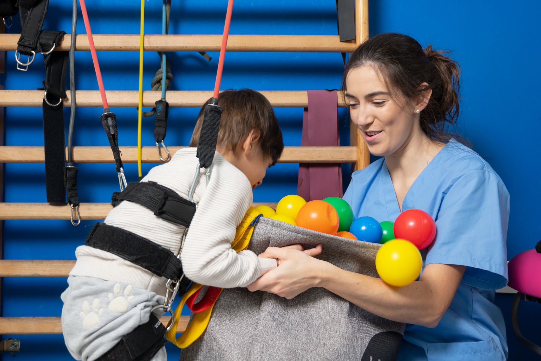
 Children with physical developmental delays will have delayed pelvic floor control as well. As you learned earlier in this blog, establishing control in positions including prone, sitting, quadruped, and standing, develops a baby’s spinal curvatures.
If children have conditions such as spina bifida or cerebral palsy, those developmental positions may be delayed or sometimes never reached.
Improper spinal curvature early in life will affect a child’s ability to attain or maintain postural positions required for voiding and defecation. This will also delay the development of the relationship between the diaphragm and pelvic floor control.
Start looking at these milestones not only from your perspective of postural control and gross motor function but also to help improve their bowel and bladder function:
- holding head up in prone
- sitting independently
- quadruped reaching and crawling
- standing independently
- walking
If a child never stands or walks, they will struggle to develop diaphragm and pelvic floor control because gravity will not challenge this system. Spending time in upright positions by using assistive devices such as standers or walkers will help develop respiratory capacity and pelvic floor control.
When you have this treatment outlook, you can help parents shift their perspectives too. Parents of children with disabilities are often overwhelmed and tired. Help them to understand the benefits of continued work in practicing and attaining developmental postural and motor skills.
 The new course, Pediatric Postural Development debuts on September 29, 2024 and focuses on the role of the pelvic floor, diaphragm, and core. This one-day course is designed to help therapists understand the development of the diaphragm and pelvic floor muscles (PFM) as they relate to core function and continence in children. Learn how to connect the ribcage, the diaphragm, and the pelvic floor for proper core activation, as well as receive instruction in anatomy and development of the diaphragm and its relationship to the pelvic floor/core. The information presented in the course applies to children who have been diagnosed with Cerebral Palsy, Down syndrome, ASD, Hypotonia, and more.
References:
- Bordoni B. The Five Diaphragms in Osteopathic Manipulative Medicine: Myofascial Relationships, Part 1. Cureus. 2020 Apr 23;12(4):e7794. doi: 10.7759/cureus.7794. PMID: 32461863; PMCID: PMC7243635.
- Kretch, K. S., Marcinowski, E. C., Hsu, Y., Koziol, N. A., Harbourne, R. T., Lobo, M. A., & Dusing, S. C. (2023). Opportunities for learning and social interaction in infant sitting: Effects of sitting support, sitting skill, and gross motor delay. Developmental Science, 26(3), e13318. https://doi.org/10.1111/desc.13318
Special thanks to Dawn for allowing The Pelvic Rehab Report to reprint her article, originally published on her website at kidsbowelbladder.com.
AUTHOR BIO:
Dawn Sandalcidi PT, RCMT, BCB-PMD
Dawn Sandalcidi is a trailblazer and leading expert in the field of pediatric pelvic floor disorders. She graduated from SUNY Upstate Medical Center in 1982 and is actively seeing patients in her clinic Physical Therapy Specialists, Centennial CO.
Dawn is a national and international speaker in the field, and she has gained so much from sharing experiences with her colleagues around the globe. In addition to lecturing internationally on pediatric bowel and bladder disorders, Dawn is also a faculty instructor at the Herman & Wallace Pelvic Rehab Institute. Additionally, she runs an online teaching and mentoring platform for parents and professionals.
In 2017, Dawn was invited to speak at the World Physical Therapy Conference in South Africa about pediatric pelvic floor dysfunction and incontinence. Dawn is also Board-Certified Biofeedback in Pelvic Muscle Dysfunction (BCB-PMD). She has also been published in the Journals of Urologic Nursing and Section of Women’s Health.
In 2018, Dawn was awarded the Elizabeth Noble Award by the American Physical Therapy Association Section on Women's Health for providing Extraordinary and Exemplary Service to the Field of Physical Therapy for Children.


Faculty member Ginger Garner PT, DPT, ATC/L is offering a new short course, The Voice and The Pelvic Floor. This course introduces foundational concepts needed to be able to use vocal techniques to treat the pelvic floor and core. Dr. Garner is a clinician, educator, and longtime advocate committed to making physical therapy services accessible, affordable, and equitable, especially for pelvic health.

When you think of pelvic health, what comes to mind? Obvious variables like the pelvic floor & girdle, abdominals and related synergists, mental health, and gut and respiratory health are probably at the top of your list.
But how often do we consider the voice as a biomarker of pelvic health? It can impact all of the variables above and more.
Historically, speech-language pathologists’ study of vocal health has stopped at the respiratory diaphragm, while physical and occupational therapists’ study of pelvic health has stopped, well, in the same place. Neither has traveled beyond that until recently.
However, there is a third diaphragm beyond the respiratory and pelvic, the laryngeal diaphragm. It’s also known as the cervical, cervico-thoracic, vocal, and/or thoracic diaphragm. The three diaphragms include:
- The Laryngeal Diaphragm - is responsible for neurological optimization of stress response and physiological control of swallowing and communication; but, it also influences vagal tone for cardiorespiratory functioning and respiratory and pelvic diaphragm functioning. It contains the muscles that are responsible for phonation, which includes intrinsic variables such as the arytenoids, but also extrinsic components which have a direct impact on the vocal fold health, such as the suprahyoid muscles.
- The Respiratory Diaphragm - is the connecting point between cephalad and caudad diaphragms and is the main muscle influencing pulmonary function. However, the respiratory diaphragm exacts a major influence on mind-body health, which goes far beyond pressure regulation of the vocal and pelvic diaphragms.
- The Pelvic Diaphragm - is the terminal end of the tri-diaphragmatic (3D) system, and can bear the brunt of trauma and impairment if dysfunction is present in the two upstream diaphragms. The pelvic diaphragm contains the levator ani, coccygeus, and related synergists, pelvic fascia, and neurovascular structures, which in turn can work with or against breathing and voicing tasks.
The diaphragms are in constant movement and none work in isolation. Together, their intersectional action provides us with the key to both internal and external biopsychosocial stability and structure of the mind-body.
The laryngeal diaphragm has a supradiaphragmatic vagal impact, while the pelvic diaphragm exacts subdiaphragmatic vagal health, chiefly through afferent and efferent input, respectively. The 10th cranial nerve lives up to its namesake, “the wandering,” as it touches each of the three diaphragms on its journey, harnessing the capacity to lessen pelvic and visceral pain, while also improving vocal quality and lung function, and changing pain, mood, and digestive function.
The mind-body interface of the 3D system has been further defined in recent years, broken down into a voluntary motor system (the one we spend all our time studying and treating), and the “emotional motor system,” and the implications are profound. Anatomists and researchers tell us that in order to generate speech, we need both motor systems to function. But specifically, our emotional motor system must first perceive safety before speech can be produced or produced well.
To get a feel for your own emotional motor system health, try this brief exercise:
- Think about a recent incident that made you feel nervous, or anxious.
- How well could you breathe?
- Talk?
- Sing or hum?
- Engage in intimacy with someone?
Not very well, right? This feeling is the fallout from your emotional motor system perceiving danger or threat. It’s what Dr. Stephen Porges means by the phrase, “neuroception.” Neuroception is the ability to detect risk - but it’s not just the ability to detect it - neuroception is the ability to accurately detect risk.
Here lies the problem:
If we cannot detect external risk or internal threat accurately, aka if our neuroception wiring is faulty, then we may move to 1 of 4 default modes for behavior: fight, flight, freeze, or fawn. If we are left in this state of reactivity, courtesy of the sympathetic nervous system, then polyvagal theory predicts we will enter into a dissociative state, termed a dorsal vagal response (DVR). The DVR drives self-preservation in severe trauma states, which can preserve life; but it is also to blame for bradycardia and left unchecked, death. Especially now post-COVID, it’s imperative that all therapists understand how to recognize, screen for, and help nurture healthy self-regulatory strategies via trauma-informed care.
We must also learn how to create a therapeutic landscape conducive to healthy neuroception, one that appeals to the “safety switch” of the emotional motor system. Establishing this healthy therapeutic alliance with our patients and clients is critical in pelvic health because the same motor system that controls the creation of sound, dictates everything associated with pelvic health, including micturition, defecation, partuition, stress response, and sexual function.
Understanding the basis of “3D” neurophysiology makes targeting the voice a perfect alternate but necessary pathway for successful comprehensive pelvic health care. The ability to create sound literally determines how we interact with the world around us, and whether or not we can do so with empathy and safety. Additionally, the success of our intervention as pelvic health therapists is also determined by the degree to which we are using a biopsychosocial model, which has long been supported as the most effective and cost-effective way to manage pain and tackle chronic disease and impairment.
What does including the voice look like in pelvic health?
For starters, the first step is evaluating the orofacial and vocal health of the patient or client. Second, and perhaps surprising to some, is the evaluation of the therapist’s own voice as a therapeutic agent. These assessments work to identify red flags that place undue stress through the downstream diaphragms and stress response system. More complex assessment can include lumbopelvic ultrasound imaging as well, which provides a more comprehensive way to individualize therapy prescription.
Assessment is essentially a 4-pronged process - The first two prongs consist of building on existing evaluation skills in respiratory, core, and pelvic floor and girdle assessment. The final two include an assessment of the physiological functioning of the orofacial area and voice and an evaluation of psychosocial determinants which would influence cranial nerve and vocal functioning. The voice is an incredible tool for improving pelvic health outcomes, if we learn how to harness its frequency and power.
Join Dr. Ginger Garner for a virtual short course, The Voice and the Pelvic Floor scheduled for October 1, 2022, to learn more.
Sources
- HOLSTEGE, G., 2016. How the Emotional Motor System Controls the Pelvic Organs. Sexual Medicine Reviews, 4(4), pp. 303-328.
- HOLSTEGE, G. and SUBRAMANIAN, H.H., 2016. Two different motor systems are needed to generate human speech. The Journal of comparative neurology, 524(8), pp. 1558-1577.
- Speer LM, Mushkbar S, Erbele T. Chronic Pelvic Pain in Women. afp. 2016;93(5):380-387.
- Miciak M, Gross DP, Joyce A. A review of the psychotherapeutic “common factors” model and its application in physical therapy: the need to consider general effects in physical therapy practice. Scand J Caring Sci. 2012;26(2):394-403. doi:10.1111/j.1471-6712.2011.00923.x
- Padoa A, McLean L, Morin M, Vandyken C. The Overactive Pelvic Floor (OPF) and Sexual Dysfunction. Part 2: Evaluation and Treatment of Sexual Dysfunction in OPF Patients. Sex Med Rev. 2021;9(1):76-92. doi:10.1016/j.sxmr.2020.04.002
- Wijma AJ, van Wilgen CP, Meeus M, Nijs J. Clinical biopsychosocial physiotherapy assessment of patients with chronic pain: The first step in pain neuroscience education. Physiother Theory Pract. 2016;32(5):368-384. doi:10.1080/09593985.2016.1194651
- Porges SW. The polyvagal perspective. BiolPsychol. 2007;74(2):116-143.
All Upcoming Continuing Education Courses

Menopause Transitions and Pelvic Rehab - Remote Course - January 17 - 18 2026
Jan 17 2026 - Jan 18 2026

Pelvic Function Level 1 - Satellite - Bethpage NY - January 24 - 25 2026- SOLD OUT
Jan 24 2026 - Jan 25 2026

Pelvic Function Level 2B - Satellite - Port St. Lucie FL - January 24 - 25 2026
Jan 24 2026 - Jan 25 2026

Pelvic Function Level 1 - Satellite - Woodbury NY - January 24 - 25 2026 - SOLD OUT
Jan 24 2026 - Jan 25 2026

Pelvic Function Level 2B - Satellite - Indianapolis IN - January 24 - 25 2026 - SOLD OUT
Jan 24 2026 - Jan 25 2026


Pelvic Function Level 2B - Satellite - St. Augustine FL - January 24 - 25 2026
Jan 24 2026 - Jan 25 2026


Pelvic Function Level 1 - Satellite - Bradenton FL - January 24 - 25 2026 - SOLD OUT
Jan 24 2026 - Jan 25 2026

Pelvic Function Level 1 - Satellite - New York NY - January 24 - 25 2026 - SOLD OUT
Jan 24 2026 - Jan 25 2026

Pelvic Function Level 1 - Satellite - Los Angeles CA - January 24 - 25 2026 - SOLD OUT
Jan 24 2026 - Jan 25 2026


Pelvic Function Level 1 - In-Person - Columbus OH - January 31 - February 1 2026
Jan 31 2026 - Feb 1 2026

Pelvic Function Level 2C - Satellite - Boston MA - January 31 - February 1 2026
Jan 31 2026 - Feb 1 2026

Pelvic Function Level 2C - Satellite - Omaha NE - January 31 - February 1 2026
Jan 31 2026 - Feb 1 2026


Pelvic Function Level 1 - Satellite - Fairfax VA - February 7 - 8 2026 - SOLD OUT
Feb 7 2026 - Feb 8 2026

Pelvic Function Level 1 - Satellite - Queens NY - February 7 - 8 2026 - SOLD OUT
Feb 7 2026 - Feb 8 2026


Pelvic Function Level 1 - Satellite - Greenwich Village NY - February 7 - 8 2026 - SOLD OUT
Feb 7 2026 - Feb 8 2026


Pelvic Function Level 2B - Satellite - Seattle WA - February 14 - 15 2026 - SOLD OUT
Feb 14 2026 - Feb 15 2026

Pelvic Function Level 2B - Satellite - Galloway NJ - February 14 - 15 2026
Feb 14 2026 - Feb 15 2026

Pelvic Function Level 2B - Satellite - Owensboro KY - February 14 - 15 2026
Feb 14 2026 - Feb 15 2026

Mobilization of Visceral Fascia: Urinary - Satellite Lab Course - Torrance CA - February 20 - 22 2026
Feb 20 2026 - Feb 22 2026

Mobilization of Visceral Fascia: The Urinary System Satellite Lab Course - Self-Hosted - February 20 - 22 2026
Feb 20 2026 - Feb 22 2026

Mobilization of Visceral Fascia: Urinary - Satellite Lab Course - Lake Stevens WA - February 20 - 22 2026
Feb 20 2026 - Feb 22 2026

Mobilization of Visceral Fascia: Urinary - Satellite Lab Course - Medford OR - February 20 - 22 2026
Feb 20 2026 - Feb 22 2026


Pelvic Function Level 1 - Satellite - Nashville TN - February 21 - 22 2026
Feb 21 2026 - Feb 22 2026
Pain Science for the Chronic Pelvic Pain Population - Remote Course - February 21 - 22 2026
Feb 21 2026 - Feb 22 2026