

From Home Care to Pelvic Health: A Journey Guided by Faith, Mentorship, and Resilience
Sometimes the most meaningful career paths aren’t the ones we plan. For Herman & Wallace faculty member Carole High Gross, PT, MS, DPT, PRPC, the road to becoming a leader in pelvic health rehabilitation was shaped by unexpected challenges, pivotal relationships, and a willingness to trust the journey even when the destination wasn’t yet visible.
We recently sat down with Carole to talk about her career, her calling, and the work that drives her. What unfolded was one of the most compelling stories of resilience and purpose we’ve heard.
A Career Built on Breadth
Carole’s career in physical therapy spans more than three decades. After earning her Master of Science in Physical Therapy from Thomas Jefferson University in 1992, she worked across nearly every clinical setting imaginable: pediatrics, aquatics, outpatient orthopedics, inpatient rehab, contract work, and home care, which she loved most. She built a deep clinical foundation long before pelvic health was on her radar.
Then life intervened.
Carole was diagnosed with breast cancer, followed by a rare chronic leukemia called hairy cell leukemia. She also lives with CIDP, a neurological condition that significantly impacted her mobility. At one point, she was using a walker, a wheelchair, and a scooter for community outings. Clinical work, at least the way she’d always done it, was no longer an option.
But Carole’s response was characteristically forward-looking: her brain was still working, so she went back for her doctorate.
Getting Back Into the Swing of Things
When Carole enrolled in her Doctor of Physical Therapy program at Arcadia University, the same institution where she’d started her undergraduate education years earlier (she lovingly calls them her “bookend university”), the transition wasn’t easy. She recalls sitting on her bed, textbooks in hand, wondering why she was putting herself through it.
But she found a way to reframe the challenge. She hadn’t forgotten how to learn. She’d simply had a very long summer. That simple mindset shift became a guiding mantra. Every time Carole faces a challenge in her health, her career, or her education, she reminds herself that she’s just getting back into the swing of things.
Walking Through the Door
As Carole neared the end of her DPT, she knew she couldn’t return to home care. She felt pulled toward something but didn’t know what it was. She describes it as trusting a GPS where someone else can see the full route, but she can only see the next turn on the screen.
Then, in a matter of days, a series of small, seemingly random events changed the trajectory of her career.
A friend convinced her to stop by a retirement party. There, she bumped into Kathy Sumner, a PT she’d worked with 20 years earlier. Kathy invited Carole to visit a pelvic health clinic she ran with Janet Whelan Drake, who Carole now works alongside as a Lead Teaching Assistant at Herman & Wallace.
When Carole walked through the clinic door, the feeling was immediate and unmistakable. She was home.
Kathy and Janet became Carole’s mentors. Weekends of hands-on training. Patients brought in for teaching opportunities. Encouragement to pursue coursework. The small-room private practice setting turned out to be the perfect environment for someone navigating mobility challenges, a place where Carole could not only survive, but thrive.
The timing was ideal. Her DPT program required a semester-long research project on a topic of interest, and Carole channeled everything into developing her Belly After Baby program for postpartum women, with Kathy and Janet guiding her every step of the way.
Eating Disorders and Pelvic Health: A Critical Connection
Today, Carole is a Pelvic Clinical Rehabilitation Specialist at Jefferson Health Lehigh Valley in Pennsylvania, where she treats patients of all genders with pelvic, bowel, bladder, and abdominal concerns. She holds her Pelvic Rehabilitation Practitioner Certification (PRPC) and serves as both an instructor and Lead Teaching Assistant at Herman & Wallace.
Her course, Eating Disorders and Pelvic Health Rehabilitation: The Role of a Rehab Professional, fills a critical gap in pelvic health education. Individuals with eating disorders frequently present with the exact symptoms pelvic rehab professionals treat every day: constipation, bloating, abdominal pain, pelvic organ prolapse, urinary dysfunction, and pelvic pain. Yet the connection between eating disorders and pelvic health is often overlooked.
As Carole explains, pelvic health providers aren’t going to diagnose or treat eating disorders, but they absolutely can and should be asking the right questions. They can observe, support, refer, and provide manual and educational tools that make a real difference in someone’s recovery journey. Sometimes, a pelvic health clinician is the first provider to notice the signs and gently guide someone toward help.
The course has received outstanding reviews, with clinicians praising its depth and Carole’s ability to connect the bigger picture, the multidisciplinary web of providers that supports individuals with eating disorders, with the specific, actionable skills pelvic health professionals can bring to the table.
Research at the International Level
Beyond Herman & Wallace, Carole serves on the Pelvic Workgroup of the International Consortium on the Ehlers-Danlos Syndromes and Hypermobility Spectrum Disorders, facilitated by the Ehlers-Danlos Society. In 2024, the workgroup published a landmark paper in PLOS ONE, a multidisciplinary, multinational effort co-creating evidence-based clinical guidelines for the management of pregnancy, birth, and postpartum recovery in individuals with hypermobile Ehlers-Danlos syndrome (hEDS) and hypermobility spectrum disorders (HSD).
The workgroup is currently finalizing a paper focused on pelvic health concerns in individuals with hEDS and HSD, with additional publications expected through 2026 and into 2027, including updates to diagnostic criteria and guidance across multiple clinical domains.
Carole is passionate about the screening role pelvic health professionals can play for hypermobility. As she describes it, asking just a few simple questions about a history of joint subluxations, dislocations, or being “super bendy” can start to connect dots that no one else has connected. Many individuals with hypermobility present with pelvic dysfunction, GI issues, chronic pain, skin changes, and temperature sensitivities. Pelvic health clinicians may be the first to notice that these seemingly unrelated issues share a common thread.
A Philosophy of Mentorship
One theme that runs through every chapter of Carole’s story is mentorship. She was mentored into pelvic health by Kathy and Janet. She was encouraged to take that first Pelvic Floor Level 1 course by people who believed in her when she wasn’t sure she believed in herself. And now, she pays it forward: mentoring new clinicians, serving as boots on the ground at satellite courses, and fostering the collaborative, family-like learning environment that she believes is the heart of what Herman & Wallace does best.
Her advice to clinicians who feel overwhelmed by the breadth of pelvic health education?
“Keep your focus on the step you’re on. Don’t look up at the full staircase. There’s no timeline. One course, one skill, one patient at a time, and before you know it, you’ll have built something incredible underneath you.”
About Carole
Carole High Gross, PT, MS, DPT, PRPC (she/her) earned her Doctorate of Physical Therapy from Arcadia University in 2015 and her Master of Science in Physical Therapy from Thomas Jefferson University in 1992. She works as a Pelvic Clinical Rehabilitation Specialist at Jefferson Health Lehigh Valley and serves as a Lead Teaching Assistant and instructor at Herman & Wallace, where she created and teaches Eating Disorders and Pelvic Health Rehabilitation: The Role of a Rehab Professional. Carole is a member of the Pelvic Workgroup of the Ehlers-Danlos International Consortium and has a special interest in working with individuals living with eating disorders and hypermobility throughout the pregnancy and postpartum journey. She is a dedicated mentor for growing pelvic professionals and focuses on team building and program development.
Learn From Carole
Ready to explore the intersection of eating disorders and pelvic health rehabilitation? Carole’s course is designed to expand your clinical lens, build your confidence in screening and observation, and equip you with practical tools to support individuals with eating disorders on their recovery journey.
Eating Disorders and Pelvic Health Rehabilitation: The Role of a Rehab Professional
Remote Course | October 4–5, 2025 | Live via Zoom
Your patients deserve comprehensive care, and you deserve the knowledge to deliver it. Register today at hermanwallace.com. Spots are limited.


A new peer-reviewed publication in the Journal of Women’s & Pelvic Health Physical Therapy expands the evidence base for non-surgical, patient-centered care.
By Rachna Mehta, PT, DPT, CIMT, OCS, PRPC, RYT 200
Herman & Wallace Faculty Member
We are thrilled to share exciting news from Herman & Wallace faculty member Rachna Mehta, PT, DPT, CIMT, OCS, PRPC, RYT 200. Rachna has co-authored a new case report published in the Journal of Women’s & Pelvic Health Physical Therapy, alongside her colleague Becky Parr, PT, DPT, DHSc, OCS, Cert. DN, CAPP-OB.
NEW PUBLICATION
Conservative Management of Rectal Prolapse: An Integrative Physical Therapy Approach
Rachna Mehta, PT, DPT, CIMT, OCS, PRPC, RYT 200 & Becky Parr, PT, DPT, DHSc, OCS, Cert. DN, CAPP-OB
Journal of Women’s & Pelvic Health Physical Therapy, January 2026
This case report highlights a conservative, integrative physical therapy approach to managing rectal prolapse using acupressure combined with traditional pelvic floor rehabilitation. Severe rectal prolapse is a condition frequently viewed as primarily surgical in nature, often leaving older adults or those who are not ideal surgical candidates with limited options.
“This work helps expand the evidence for non-surgical, patient-centered options, particularly for older adults or those who are not ideal surgical candidates.”
— Rachna Mehta
By documenting this integrative case, Rachna and her co-author have contributed meaningfully to the growing body of evidence supporting complementary and conservative approaches within pelvic health physical therapy. This publication reflects the kind of whole-person, evidence-informed care that Rachna has championed throughout her career.
This publication is a natural extension of Rachna’s clinical expertise and her ongoing work bridging Traditional Chinese Medicine principles including acupressure and meridian theory with contemporary pelvic floor rehabilitation. Her Herman & Wallace course, Acupressure for Optimal Pelvic Health, has introduced countless clinicians to this integrative framework.
About the Author
Rachna Mehta, PT, DPT, CIMT, OCS, PRPC, RYT 200
Herman & Wallace Faculty Member · Columbia University, DPT · Board-Certified in Orthopedics · Certified Pelvic Rehab Practitioner · Registered Yoga Teacher
Rachna graduated from Columbia University with her Doctor of Physical Therapy degree and has spent over 15 years practicing in outpatient hospital and private practice settings with a dual focus on orthopedics and pelvic health. She was instrumental in founding one of the first Women’s Health Programs in an outpatient orthopedic clinic setting in Mercer County, New Jersey in 2009. Rachna also owns TeachPhysio, a PT education and management consulting company. Her clinical approach blends traditional physical therapy with holistic practices that address the whole person physically, mentally, emotionally, and spiritually.
Learn from Rachna
Explore Rachna’s course Acupressure for Optimal Pelvic Health and bring integrative acupressure into your pelvic health practice.
View Rachna’s Courses at Herman & Wallace →


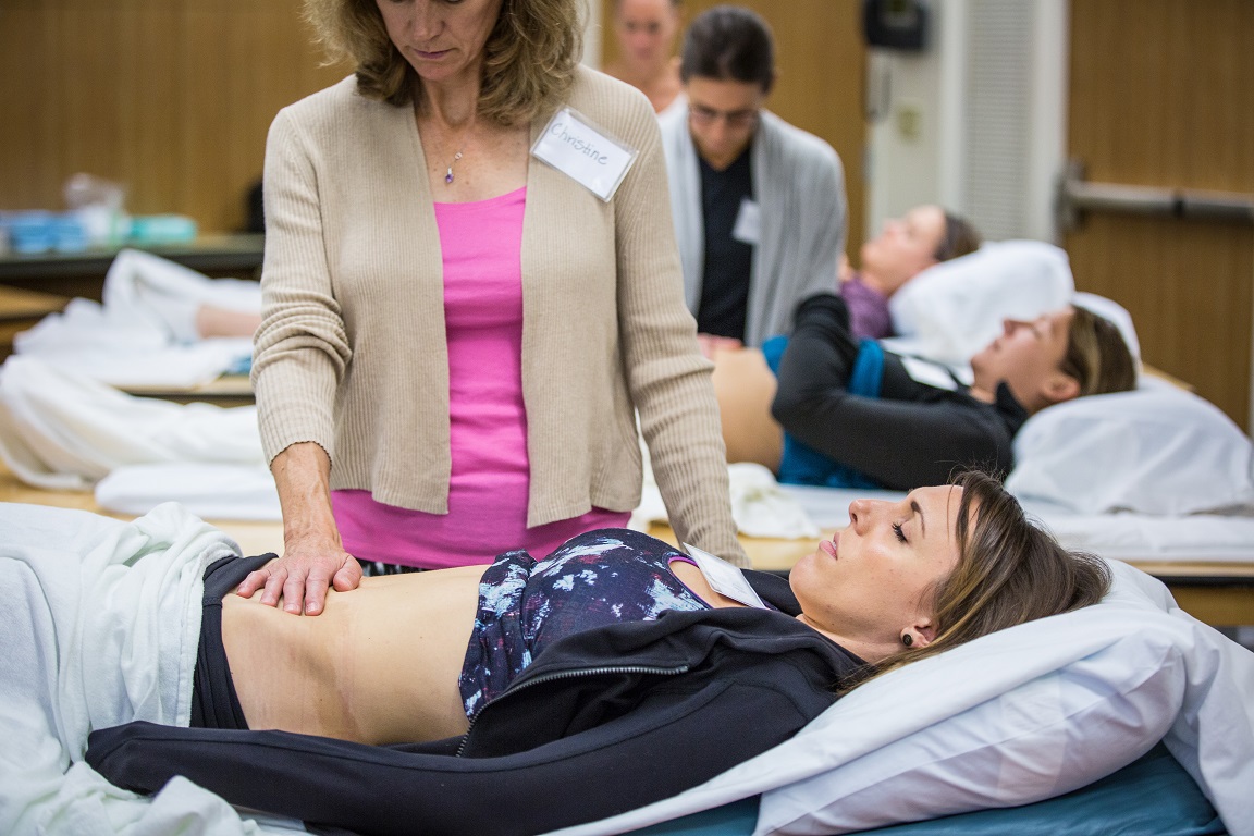
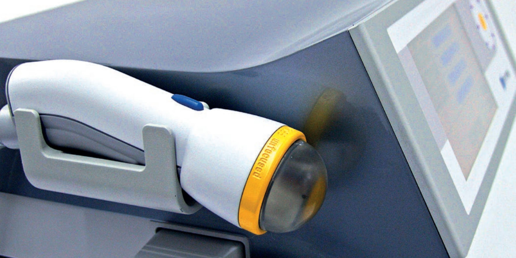
Rehabilitative ultrasound imaging is a clinical tool that can change the way you practice. I have often shared with other clinicians how much the use of ultrasound imaging has influenced how I approach patients with chronic back or sacroiliac joint pain. Using ultrasound imaging allows for a way to assess the deeper core muscles, which may be more difficult to palpate on some individuals. Being able to view the activation in these muscles can inform the therapist whether the patient is properly activating their core or relying on a less ideal strategy.
Seeing the Core Muscles That Are Hard to Reach
One of the most valuable things about rehabilitative ultrasound imaging in pelvic health is what it shows us about the deeper core muscles. These are muscles that conventional palpation simply cannot reach reliably in every patient. With ultrasound imaging, we can observe in real time whether a patient is using a proper activation strategy or compensating in a way that looks adequate on the surface but is not providing the stability they need.
That kind of information changes treatment. It gives both the clinician and the patient something concrete to work with, and it often unlocks progress that had stalled.
A Game Changer for Incontinence and Prolapse
The use of rehabilitative ultrasound imaging has also been a game changer in treating incontinence and prolapse patients. Not only does it enable me to view activation in the pelvic floor, but also the supportive function of the pelvic floor. For some patients, that supportive function is exactly what has been missing. Being able to show them what is happening in their own body, in real time, is often what finally moves treatment forward.
When Ultrasound Made All the Difference: A Patient Story
I recently began working with a patient who is a semi-professional athlete. She was 14 months postpartum and seeking care for prolapse symptoms and discomfort. This patient understood the importance of the pelvic floor and had sought out pelvic floor rehab immediately following delivery. She was approved to return to exercise and at the sixth minute of activity felt a prolapse occur.
After returning and continuing with pelvic health therapy, she still was not seeing progress with respect to her symptoms. There was real pressure mounting because she had qualified for an international level event in her sport that was six months away.
When I evaluated her, I identified that she was able to activate her pelvic floor while in a supine position, but not when standing or during a motor task. Using rehabilitative ultrasound imaging allowed her to visualize what it felt like to do a proper contraction while in standing. This was transformative. It helped her learn to engage her pelvic floor in a weight bearing position, which improved the supportive function of the pelvic floor and allowed her to begin engaging it during her sports activity.
It did take time and a lot of practice. But the addition of ultrasound imaging was what made the difference between her earlier attempts at pelvic rehab and this course of treatment.
About the Instructor: Allison Ariail, PT, DPT, CLT-LANA, BCB-PMD, PRPC
Allison Ariail, PT, DPT, CLT-LANA, BCB-PMD, PRPC is the creator of the Rehabilitative Ultrasound Imaging courses at Herman & Wallace and currently serves as Director of Education. A physical therapist since 1999, Allison holds a Doctor of Physical Therapy from Boston University. She is board certified by the Lymphology Association of North America (2011), board certified in Biofeedback Pelvic Muscle Dysfunction (2012), and earned her Pelvic Rehabilitation Practitioner Certification in 2014. She is a published researcher, a co-author in Healing in Urology, and a nationally recognized lecturer on ultrasound imaging, lymphedema, and pelvic floor dysfunction. Allison practices at Inspire Physical Therapy and Wellness in the Denver metro area, treating men, women, and children across a wide range of pelvic health conditions.
Join Us in Edmond, Oklahoma: April 17 to 19, 2026
This three-day course covers transabdominal viewing of the pelvic floor, abdominal wall, and spinal muscles as well as transperineal imaging that allows us to view the supportive function of the pelvic floor. Topics include:
- Real-time imaging of the transverse abdominals, rectus abdominis, deep multifidus, levator ani, bladder, bladder neck, urethra, and vagina
- Transabdominal and transperineal viewing methods
- Hands-on lab time with ultrasound machines provided by course sponsors
- Clinical application for lumbopelvic pain, pelvic organ prolapse, and urinary incontinence
You will love learning to use this clinical tool and seeing the changes it makes for your patients. Register for Rehabilitative Ultrasound Imaging: Pelvic Health and Orthopedic Topics in Edmond, OK at hermanwallace.com.

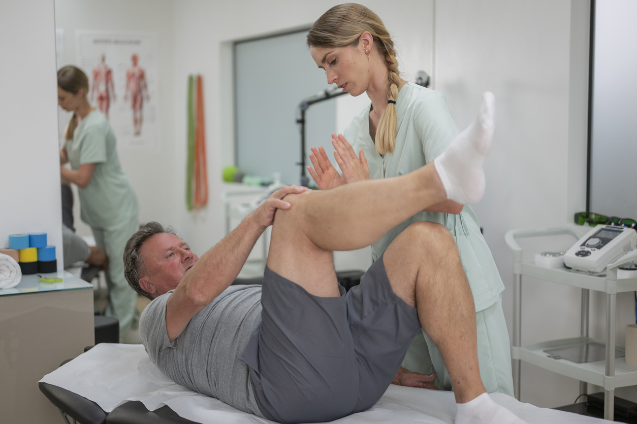
Pelvic health physical therapy sits at the intersection of multiple body systems: musculoskeletal, neuromuscular, gastrointestinal, urologic, reproductive, and psychological. Yet one critical piece is consistently underemphasized in clinical training: pharmacology.
As pelvic health providers, we routinely treat patients who are taking medications that directly influence bladder function, bowel motility, hormonal balance, tissue integrity, pain perception, sexual function, and autonomic regulation. If we are not confident in our understanding of those medications, we risk missing key contributors to our patients' symptoms — or worse, misinterpreting clinical presentation altogether.
Expanding pharmacologic literacy for pelvic health providers isn't optional anymore. It's essential.
Medications Influence the Pelvic Floor More Than We Think
Consider how commonly our patients are prescribed medications like these:
- Anticholinergics for overactive bladder
- Beta-3 agonists for urinary urgency
- Hormonal contraceptives or menopausal hormone therapy
- SSRIs and SNRIs
- Muscle relaxants
- Opioids
- Laxatives or stool softeners
Each of these medications can alter tissue quality, muscle tone, coordination, libido, arousal, bowel patterns, or pain processing. For example, oxybutynin (an anticholinergic) may reduce bladder urgency but contribute to constipation — which in turn increases pelvic floor strain. Hormonal changes driven by oral contraceptives or menopause can affect collagen integrity, vaginal tissue health, and load tolerance. Antidepressants may improve mood while simultaneously influencing sexual function or arousal.
When we assess biomechanics without considering pharmacology for pelvic health, we are seeing only part of the picture.
Medication Side Effects Can Mimic or Exacerbate Dysfunction
Patients frequently present with symptoms such as:
- Constipation or bowel irregularity
- Urinary retention, urgency, or frequency
- Sexual dysfunction or decreased arousal
- Vaginal dryness or tissue irritation
- Fatigue or dizziness affecting exercise tolerance
How often are these attributed solely to pelvic floor dysfunction when medication side effects may be a primary contributor? Understanding pharmacodynamics and pharmacokinetics allows us to recognize red flags early, identify medication-induced symptoms, modify exercise dosing appropriately, and collaborate more effectively with prescribing providers. This elevates our clinical reasoning from symptom management to genuinely comprehensive pelvic health care.
Interdisciplinary Collaboration Starts with Pharmacologic Fluency
Pelvic health PTs frequently collaborate with OB-GYNs, urogynecologists, urologists, gastroenterologists, pain specialists, and primary care providers. When we understand the indications for common pelvic medications — their mechanisms of action, contraindications, and side effect profiles — we can communicate clearly, advocate effectively for our patients, and participate meaningfully in care decisions.
This isn't about prescribing. It's about being an informed provider within a multidisciplinary team. Pharmacologic literacy is what allows pelvic health providers to show up at that table as true clinical partners.
Meet the Faculty: Kristina Koch, PT, DPT, CLT, PCES
Kristina Koch, PT, DPT, CLT, PCES is a board-certified specialist in women's health physical therapy and the creator of this course. With over two decades of clinical experience treating pelvic floor dysfunction across all genders and ages, Kristina brings unparalleled depth to this subject. She earned her doctorate from The College of St. Scholastica in 2021 and currently practices in Colorado Springs, CO, where she also serves as a guest lecturer for graduate PT students at Regis University in Denver and provides educational sessions for medical providers and community groups. Kristina has developed this course because she believes pharmacologic literacy is a professional responsibility — not just a clinical nice-to-have.
Invest in the Provider You're Becoming
Pelvic health is evolving rapidly. Our patients are complex. Our role is expanding. To practice at a high level, we must think beyond muscles and manual therapy — we must understand the biochemical and systemic influences that shape pelvic function, including the medications our patients take every day.
If you are ready to deepen your expertise, improve patient outcomes, and elevate your confidence in pharmacology for pelvic health, this is the course you've been waiting for.
Pharmacologic Considerations for the Pelvic Health Provider
Remote Course — Saturday, April 4, 2026 | 8:00 AM Pacific | Live via Zoom
This one-day remote course is designed specifically for pelvic health rehabilitation professionals and covers:
- Urologic medications and their effects on pelvic floor function
- Gastrointestinal pharmacology
- Hormonal therapies including gender-affirming care medications
- Pain medications and their impact on neuromuscular function
- Psychotropic medications and pelvic health
- Medication side effects that influence pelvic function
- Clinical decision-making and interdisciplinary communication
- Non-medication treatment alternatives
Your patients deserve comprehensive care, and you deserve the knowledge to deliver it. Register today at hermanwallace.com — spots are limited.

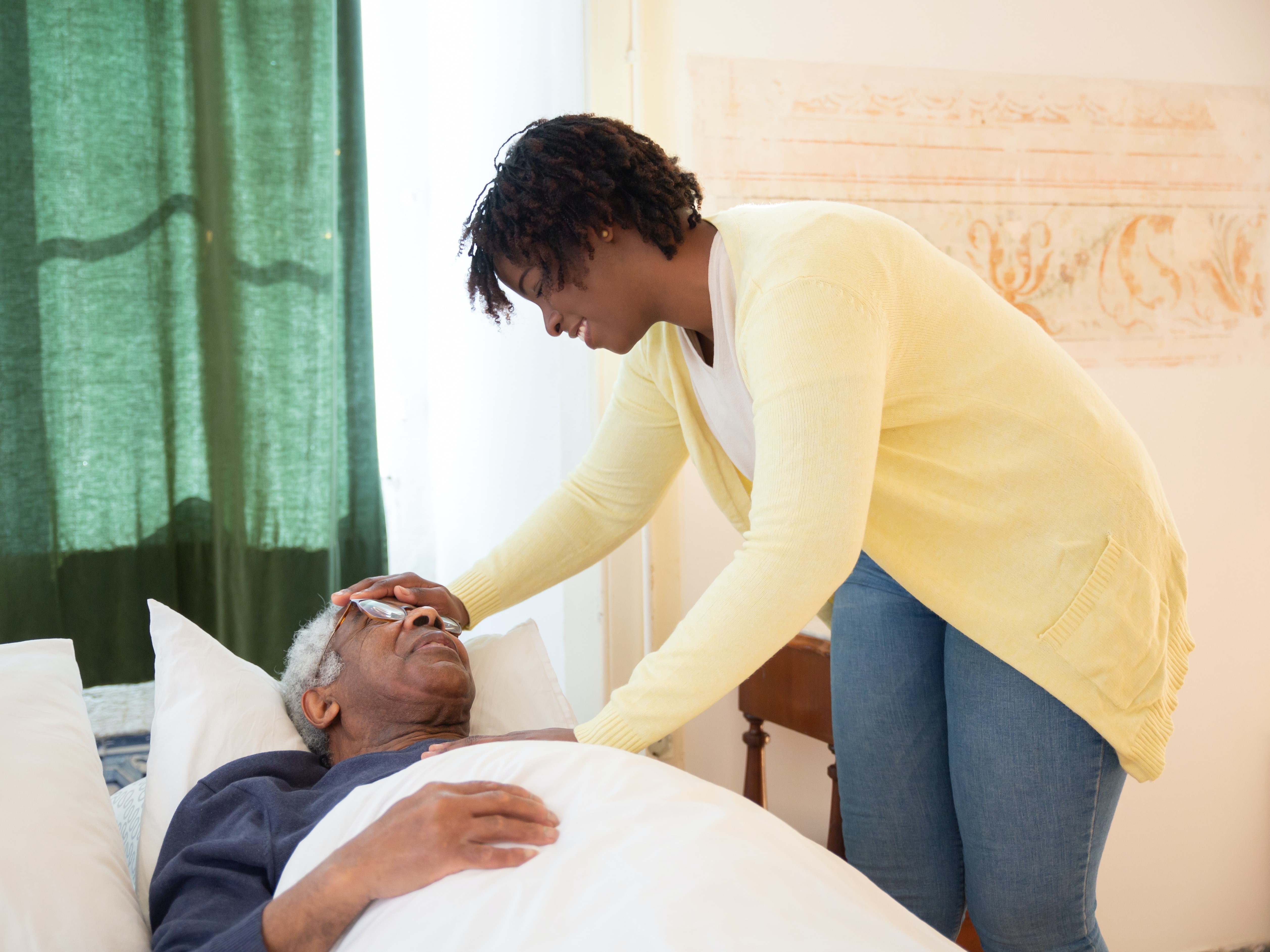
Pelvic floor rehabilitation plays an important role in helping men prepare for and recover from treatment for prostate and other pelvic cancers. Targeted pelvic rehabilitation interventions can address urinary continence, bowel function, pelvic pain, and sexual health during both preoperative preparation and post treatment recovery.
Men undergoing treatment for pelvic cancers, including prostate, bladder, penile, and testicular cancers, frequently experience changes in pelvic floor function that affect urinary, bowel, and sexual health. Surgical procedures such as radical prostatectomy, radiation therapy, chemotherapy, and reconstructive surgeries can alter pelvic floor muscle coordination, nerve signaling, and connective tissue mobility. As survivorship improves, clinicians are increasingly recognizing the role of pelvic rehabilitation in addressing these functional changes.
Pelvic floor therapy can be introduced both before treatment and during the recovery period. Preoperative pelvic floor training, often referred to as pelvic floor prehabilitation, allows patients to learn correct muscle activation and coordination prior to surgery. For individuals undergoing prostatectomy, this preparation may support earlier return of urinary continence and improve adherence to postoperative rehabilitation programs.
Following cancer treatment, patients may present with a range of pelvic health concerns including urinary incontinence, bowel dysfunction, pelvic pain, sexual dysfunction, and reduced physical confidence. In clinical practice, these symptoms often overlap and may be influenced by muscle weakness, impaired coordination, neural disruption, and tissue changes such as fibrosis.
For pelvic health clinicians, understanding the role of rehabilitation across the cancer care continuum is essential. Targeted pelvic floor interventions can support recovery of bladder control, bowel regulation, and sexual function while also addressing movement patterns, scar tissue mobility, and patient education during survivorship.
Pelvic Floor Prehabilitation Before Prostate Surgery
Preoperative pelvic floor muscle training is increasingly recommended for patients preparing to undergo radical prostatectomy. The goal of prehabilitation is to help patients identify and properly activate the pelvic floor muscles prior to surgery.
In clinical practice, many patients struggle to isolate the pelvic floor muscles without guided instruction. Teaching correct muscle activation before surgery allows patients to develop familiarity with the exercises that will be used during postoperative rehabilitation. This preparation may also improve adherence to home exercise programs during the recovery phase.
Evidence suggests that patients who perform pelvic floor muscle training prior to prostate surgery may experience faster recovery of urinary continence compared with those who begin training only after surgery. Prehabilitation also provides an opportunity to educate patients about expected postoperative changes, bladder management strategies, and activity progression during early recovery.
For many patients, this early education helps reduce anxiety and allows them to approach surgery with a clearer understanding of the rehabilitation process.
The Role of Pelvic Floor Rehabilitation After Treatment
Pelvic rehabilitation is widely recognized as an important component of recovery following prostate cancer treatment. Structured pelvic floor muscle training programs have consistently demonstrated improvements in urinary continence after radical prostatectomy.
Programs that include supervision by a pelvic health clinician often produce stronger outcomes than unsupervised exercise programs. In addition to strengthening the pelvic floor muscles, rehabilitation may address motor control, endurance, and coordination required for functional bladder support.
Accurate muscle activation is an important part of successful rehabilitation. Techniques such as biofeedback, external or internal palpation, and clinician guided instruction can help ensure that patients are engaging the pelvic floor muscles effectively during training.
Radiation therapy directed toward the pelvis can also contribute to long term changes in pelvic floor muscle function. Patients who receive pelvic radiation may develop reduced muscle endurance, altered neuromuscular coordination, and tissue changes related to fibrosis. These factors may contribute to urinary symptoms, bowel dysfunction, or pelvic pain. Rehabilitation strategies may therefore include interventions focused on muscle retraining, tissue mobility, and symptom management.
Addressing Bowel Dysfunction After Colorectal Cancer Treatment
Pelvic rehabilitation may also play an important role in the management of bowel symptoms following colorectal cancer surgery. Patients who undergo low anterior resection may develop a constellation of symptoms commonly referred to as low anterior resection syndrome. These symptoms may include bowel urgency, stool clustering, and reduced bowel control.
Targeted pelvic floor rehabilitation programs that include strengthening exercises, coordination training, and biofeedback have been shown to improve bowel function and quality of life in this patient population.
From a clinical perspective, improving coordination between pelvic floor muscles and abdominal pressure regulation is often a key component of treatment. Education regarding bowel habits, defecation mechanics, and behavioral strategies may also support symptom management.
Supporting Sexual Function During Recovery
Sexual health is another domain in which pelvic rehabilitation can contribute to recovery following pelvic cancer treatment. Pelvic floor muscle training has been shown to support improvements in erectile function, particularly when incorporated into structured rehabilitation programs before or after prostate surgery.
In addition to strengthening muscles involved in erectile function, pelvic rehabilitation may address pelvic pain, scar tissue sensitivity, and muscle coordination impairments that can contribute to discomfort during sexual activity.
Because sexual health concerns often involve both physical and psychosocial components, collaboration with urology, sexual medicine, and mental health professionals can provide comprehensive support for patients navigating these changes during survivorship.
The Expanding Role of Pelvic Rehabilitation in Cancer Survivorship
As survival rates continue to improve for men with pelvic cancers, greater attention is being placed on functional recovery and quality of life following treatment. Pelvic rehabilitation provides clinicians with an opportunity to address bladder control, bowel function, sexual health, and pelvic pain in a structured and evidence informed way.
For many patients, these symptoms are not simply unavoidable consequences of cancer treatment. With appropriate rehabilitation strategies and coordinated care, meaningful improvements in daily function and overall wellbeing are often achievable.
Clinical Takeaways for Pelvic Health Clinicians
Pelvic rehabilitation plays an important role in supporting functional recovery for male pelvic cancer survivors. Clinicians working with this population may consider the following points.
Urinary incontinence is common following prostatectomy, and pelvic floor muscle training can support recovery of bladder control.
Preoperative pelvic floor training can help patients identify and activate the pelvic floor muscles prior to surgery, which may facilitate postoperative rehabilitation.
Radiation therapy may contribute to changes in pelvic floor muscle endurance, neuromuscular coordination, and connective tissue mobility that influence pelvic function.
Patients undergoing colorectal cancer surgery may develop bowel dysfunction that can respond to pelvic floor rehabilitation interventions.
Pelvic floor muscle training may also support recovery of erectile function and improve sexual health outcomes following prostate cancer treatment.
Collaboration among pelvic health clinicians, urologists, oncologists, and other specialists can improve patient outcomes during cancer survivorship.
Frequently Asked Questions About Pelvic Rehabilitation After Pelvic Cancer
What is pelvic floor therapy after prostate surgery?
Pelvic floor therapy focuses on restoring bladder control, improving pelvic floor muscle coordination, and supporting recovery of sexual function following prostate surgery.
When should pelvic floor therapy begin for prostate cancer patients?
Pelvic floor muscle training may begin prior to surgery as part of prehabilitation and continue after surgery during recovery.
Can pelvic rehabilitation support erectile function recovery?
Pelvic floor muscle training may support erectile function by improving muscle strength and coordination involved in sexual performance.
Is pelvic rehabilitation helpful for bowel dysfunction after colorectal cancer surgery?
Yes. Pelvic rehabilitation can help improve bowel control and coordination for patients experiencing symptoms associated with low anterior resection syndrome.
Who may benefit from pelvic cancer survivorship rehabilitation?
Men treated for prostate, bladder, colorectal, penile, or testicular cancers may benefit from pelvic rehabilitation to address bladder, bowel, sexual, and pelvic pain concerns.
Key Research References
Jones H, et al. Rehabilitation strategies for low anterior resection syndrome following colorectal cancer surgery. Colorectal Dis. 2024.
Kim HJ, Oh SY. Pelvic floor rehabilitation for bowel dysfunction after colorectal cancer surgery. Ann Coloproctol. 2023.
Anderson CA, Omar MI, Campbell SE, et al. Conservative management for postprostatectomy urinary incontinence. Cochrane Database Syst Rev. 2015.
Fernández RA, et al. Pelvic floor muscle training after radical prostatectomy improves urinary continence recovery. Urology. 2015.
Ribeiro LH, et al. Pelvic floor muscle morphology and function after pelvic radiation therapy. Neurourol Urodyn. 2021.
Centemero A, Rigatti L, Giraudo D, et al. Preoperative pelvic floor muscle exercise for early continence after radical prostatectomy. Eur Urol. 2010.
Milios JE, Ackland TR, Green DJ. Pelvic floor muscle training in radical prostatectomy. BMC Urol. 2019.
Dorey G, Speakman M, Feneley R, et al. Pelvic floor exercises for erectile dysfunction. BJU Int. 2005.
Want to learn more about Oncology of the Pelvic Floor?
Recommended Courses: Oncology of the Pelvic Floor Level 1 & Oncology of the Pelvic Floor Level 2A
"Oncology of the Pelvic Floor Level 2A" with Nicole L Dugan,PT,DPT,MSOD, CLT-LANA, WCS
Upcoming Remote Course: April 18-19, 2026 Oncology of the Pelvic Floor Level 2A


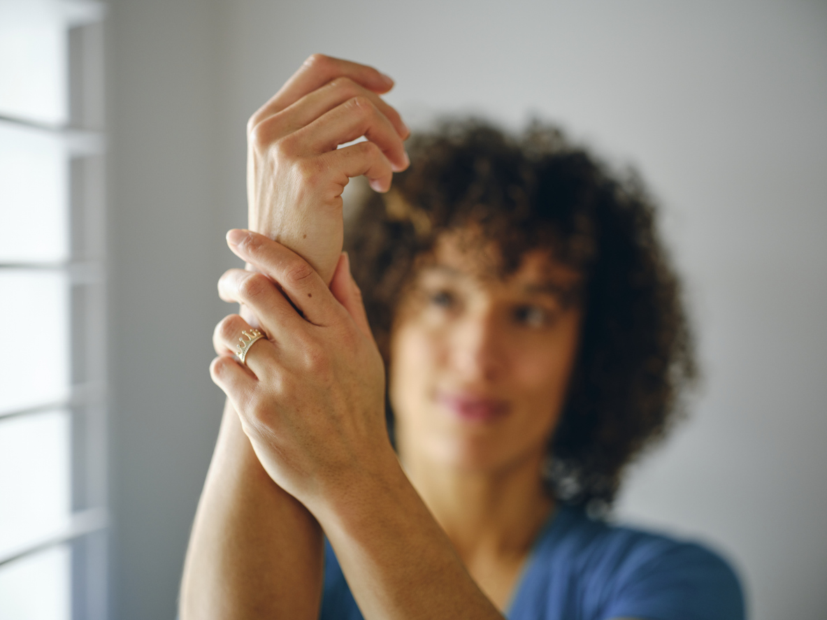
Pain shows up in almost every pelvic health plan of care, but a lot of providers were never actually trained to teach pain in a way that changes outcomes. We learn how to assess tissues, strength, tone, and pathology, but pain is not a simple “damage meter.” It’s a protective output of the nervous system, shaped by context, perceived threat, inflammation, prior experiences, hormones, and learned responses. When we skip pain science education (or keep it vague), patients often stay fearful, hypervigilant, and stuck, especially when imaging is normal or symptoms don’t “match” what we see.
Endometriosis, among other pelvic pain conditions, is one of the clearest examples of why this matters. Endometriosis is characterized by lesions containing endometrium-like epithelium and stroma that develop outside the uterus and are biologically distinct from normal uterine endometrium. Endo is a real inflammatory disease with lesions that can be found on the bowel, bladder, ureters, abdominal wall, and peritoneum commonly. And yet pain severity doesn’t reliably correlate with lesion size, number, or location. Some patients with extensive disease report minimal pain, while others with smaller disease experience life-altering symptoms. Pain science helps us explain that gap: the nervous system can become sensitized over time, turning up the volume on danger signals even when tissues are stable or after the primary driver has been addressed.
Clinically, one of the most important skills is being able to distinguish peripheral pain generators from sensitization. Peripheral drivers include things like active lesions/inflammation, adhesions, pelvic floor overactivity, tissue irritation, and organ-specific contributors. Sensitization shows up when pain persists beyond expected healing, spreads, becomes disproportionate to findings, or is paired with hypervigilance, fear-avoidance, and strong symptom reactivity to stress, sleep disruption, and attention. In pelvic health, cross-talk between organs adds another layer, bladder, bowel, uterus, and pelvic floor can share neural pathways, so symptoms don’t always point neatly to the true source.
This is where pain science education becomes a part of treatment. For endometriosis, an example of pain science education starting point could be: “Endo can absolutely create pain through inflammation and lesion activity, but pain isn’t always a direct reflection of how much disease is present. Over time, your nervous system can become extra protective, like an alarm system that’s gotten too sensitive. That doesn’t mean the pain is in your head. It means your pain IS real, and your nervous system is amplifying signals. The good news is the system can be retrained through the right combination of medical care, pelvic rehab, graded exposure, and nervous system regulation.”
When providers can explain pain clearly, patients stop interpreting every symptom spike as damage. They become more confident with movement, more consistent with rehab, and more resilient during flares. And that’s why pain science education is so important, because with endometriosis, IC/PBS, prostatitis, IBS, vaginismus/dyspareunia, and primary dysmenorrhea, your hands matter, but what you do with your words can be the turning point.
If you’re treating endometriosis, or any chronic pelvic pain condition, and you’re not sure whether you’re addressing the “spark” (peripheral drivers) versus the “fuel” (sensitization), this Pain Science class is designed to make that clinical reasoning practical, teachable, and immediately usable in your sessions.
Dr. Tara Sullivan, PT, DPT, PRPC, WCS, IF Sexual Medicine in Pelvic Rehabilitation - Remote Course - March 14-15 2026
Learn More: Sexual Medicine in Pelvic Rehab March 14-15, 2026


Anorectal balloon catheter training is one of the most underrated but helpful treatments for people with pelvic floor symptoms related to bowel dysfunction. This is a tool that many clinicians don’t know about or are afraid to initiate with their clients. Clinicians wonder if clients will be receptive, how to use an anorectal balloon catheter efficiently, and frequently wonder what cases are appropriate for this specific modality. Anorectal balloon catheter training is a versatile treatment helping patients with pelvic floor conditions that stem from hyposensitivity or hypersensitivity in the rectal canal.
Rehab clinicians can use anorectal balloon catheters to help with defecation training, anorectal sensory training, coordination training, and resistance training that can improve symptoms for individuals with fecal incontinence, fecal urgency, and chronic constipation as well as other colorectal diagnoses. This modality can be used to improve the coordination between the pelvic floor muscles and the abdominal muscles to assist in defecation training. It also can help a patient learn what the urge to have a bowel movement should feel like, especially if they have altered sensation in the anal canal.
An anorectal balloon is a form of biofeedback to use with pelvic floor patients. During treatment, an anorectal balloon is placed in the rectal canal. The balloon can hold 400 mL but filling volumes are typically much lower. The balloon is then filled with air and the amount of air is altered in order to help retrain sensation in the anorectal area. Before implementing this treatment technique in a patient’s plan of care, there are a few steps a rehabilitation provider should take.
First, patients should be screened to make sure they are good candidates for this treatment. This includes internal muscle assessment of the rectal canal prior to implementing training. Detailed patient education on the purpose and procedure of training with an anorectal balloon catheter should be provided. Patients may have some experience with anorectal manometry and may need their therapist to differentiate how manometry testing is for assessment purposes, but balloon training is a biofeedback tool.
Once this treatment is decided upon, the therapist will begin by getting some baseline measurements. These include the first feeling of sensation of the balloon filling, the first urge to defecate, and then their maximum tolerance. These baselines give a provider information on how to proceed with treatment. It is helpful to have norms readily available to be able to compare your patient’s readings to. Caution should be taken when working with patients who have had lower bowel surgeries and pediatric patients, avoiding maximum values beyond a certain value.
With proper consideration of the baseline measurements of sensation levels, a treatment plan can be developed with the use of anorectal balloon training to improve sensation and awareness in the anorectal area. Sensation is trained via inflations and deflations of the balloon to assist in feedback to allow the patient to recognize what normal range values feel like.
Anorectal Balloon Catheters - Intro and Practical Application is a mini-course offered by Herman & Wallace to help providers feel comfortable screening patients for their eligibility for this intervention. The course will assist in helping practitioners to feel confident in providing this treatment with appropriate patients. This class is built with treatment in mind, and intended for therapists who have some exposure to the concept of anorectal assessment and treatment but want to learn more ways to apply this technique to their clients. This class includes didactic information and hands-on lab practice in the privacy of participant’s own space, to help bring this skill to their clinical practice. The next offering of this course is:
https://hermanwallace.com/continuing-education-courses/anorectal-balloon-catheters/


Abdominal bloating and distension are common symptoms reported in pelvic health practice. While many individuals experience occasional bloating that resolves without intervention, persistent or long-standing distension can significantly impact quality of life. Patients often report discomfort, sleep disruption, dietary restrictions, and frustration when symptoms persist without clear answers.
One condition associated with these symptoms is abdomino-phrenic dyssynergia, a disorder involving a paradoxical relationship between the diaphragm and abdominal wall.
Under normal conditions, when intraluminal gas increases in the gastrointestinal tract, the body responds with a coordinated pattern:
- the diaphragm relaxes, and
- the abdominal wall contracts
This response helps maintain abdominal shape and pressure regulation.
However, in abdomino-phrenic dyssynergia, the opposite pattern occurs. The diaphragm contracts downward while the abdominal musculature relaxes, leading to visible abdominal distension and discomfort. Dysfunction of the pelvic floor is also frequently associated with this condition, reinforcing the importance of a comprehensive evaluation of the entire pressure management system.
Traditional management strategies include biofeedback therapy and breathing retraining, both aimed at restoring appropriate neuromuscular coordination.
A Clinical Case Example
In our clinic, we are seeing an increasing number of referrals for patients diagnosed with abdomino-phrenic dyssynergia. One recent patient illustrates how breathing mechanics and musculoskeletal restrictions can contribute to these symptoms.
The patient was a 72-year-old female with a long-standing history of abdominal bloating and distension.
She reported:
- Bloating and abdominal distension throughout the day, worsening toward evening
- Limiting evening food intake due to abdominal discomfort and “tightness”
- Pain rated 3–5/10 in the morning, increasing to 8/10 by late evening
- Difficulty sleeping due to the abdomen feeling “hard and tight” at bedtime
Examination Findings
Physical examination revealed several contributing factors:
- Significant tightness in the posterior chain and erector spinae in the thoracic and lumbar regions
- Reduced thoracic rotation and mobility
- Connective tissue restrictions in the upper abdominal quadrants, especially the epigastric region and inferior rib cage
- Decreased lower rib cage mobility
- Difficulty producing a prolonged or forceful exhale
- Reduced ability to relax the pelvic floor following contraction
These findings highlighted the interaction between breathing mechanics, rib cage mobility, myofascial restrictions, and pelvic floor coordination.
Treatment Approach
Treatment included a multi-system approach addressing breathing, mobility, and neuromuscular coordination.
Interventions included:
- Biofeedback therapy
- Visceral mobilization techniques
- Thoracic spine and rib joint mobilizations
- Soft tissue techniques, including gentle diaphragm release
- Breathing retraining
- Techniques focused on pelvic floor relaxation
The patient completed nine treatment sessions, combined with a structured home maintenance program that she followed consistently.
Outcomes
By the end of treatment, the patient reported:
- 70% overall improvement in symptoms
- Ability to eat evening meals without discomfort
- Restful sleep through the night without abdominal tightness
This case highlights how restoring efficient breathing mechanics and rib cage mobility can significantly influence abdominal pressure regulation, pelvic floor function, and patient comfort.
Why Breathing Matters for Pelvic and Orthopedic Therapists
Breathing is far more than a respiratory function. The diaphragm plays a central role in:
- pressure regulation
- core stability
- pelvic floor coordination
- movement efficiency
Understanding how breathing integrates with the musculoskeletal system can significantly expand a clinician’s ability to address persistent symptoms that may otherwise be overlooked.
In the course Breathing and the Diaphragm: Pelvic and Orthopedic Therapists, we explore these relationships in depth and provide clinicians with practical tools to assess and treat dysfunctional breathing patterns.
Participants will learn how to:
- Explain normal diaphragmatic breathing and the role of the internal and external oblique musculature
- Assess and treat dysfunctional breathing patterns including chest, abdominal, and paradoxical breathing
- Understand the role of intra-abdominal pressure (IAP) in spinal stability
- Apply the concept of regional interdependence in patients with pelvic or back pain
- Recognize how postural patterns influence diaphragm and pelvic floor function
- Identify myofascial contributors to dysfunctional breathing and apply appropriate treatment techniques
- Perform rib and thoracic spine mobilizations to improve respiratory mechanics
- Develop exercise progressions for breathing retraining in clinic and home programs
- Integrate diaphragmatic breathing strategies into athletic rehabilitation
Understanding the relationship between breathing mechanics, mobility, and pelvic floor function allows clinicians to address dysfunction from a more integrated perspective and can lead to meaningful improvements in patient outcomes.
Aparna Rajagopal, PT, MHS, WCS, PRPC, Capp-OB Certified is the lead therapist at Henry Ford Macomb Hospital's pelvic dysfunction program, where she treats pelvic rehab patients and consults with the sports therapy team. Her interest in treating peripartum patients and athletes allowed her to recognize the role that breathing plays in pelvic dysfunction.
Leeann Taptich DPT, SCS, MTC, CSCS leads the Sports Physical Therapy team at Henry Ford Macomb Hospital where she mentors a team of therapists. She also works very closely with the pelvic team at the hospital which gives her a very unique perspective of the athlete.
Aparna and Leeann co-authored the course, Breathing and the Diaphragm: Pelvic and Orthopedic Therapists, which helps clinicians understand breathing mechanics and their relationship to the pelvic floor.

Course Dates: March 14, 2026
Price: $450
Experience Level: Beginner
Contact Hours: 13.5
Description: This remote course is an integrated approach where participants will learn how the diaphragm, breathing, and the abdominals can affect core and postural stability through intra-abdominal pressure changes while looking at structures from the glottis and the cervical region to the pelvic floor.
This course includes assessment and treatment of the barriers by addressing thoracic spine articulation and rib cage abnormalities in the fascial system of muscles related to breathing and the diaphragm. Instructed techniques are applicable to patients who present with Diastasis Rectus Abdominis, pelvic pain, incontinence, and prolapse, as well as cervical, thoracic, scapular, and lumbar pain.

I recently evaluated a 75 y.o patient who presented with significant urinary urgency and frequency, voiding approximately every hour. She reported disrupted sleep due to nocturia, stating, “I can’t sleep at night because I keep getting up to go to the bathroom. They gave me medication to help me sleep, but it doesn’t work.”
Over the course of the visit, it became clear that she was also experiencing chronic anxiety. Anxiety permeated multiple aspects of her daily life, she worried about day-to-day events as well as events in the future. She reported that her urinary symptoms worsened during periods of heightened anxiety, and she had difficulty relaxing both her body and mind.
My initial clinical focus was nervous system regulation. I guided her to sit back comfortably and take several gentle breaths, emphasizing a prolonged exhalation with an audible sigh. She was instructed to consciously release tension throughout her body while maintaining attention on her breath. After only a few breaths, she smiled and reported that she already felt calmer.
In addition to a home program that included diaphragmatic breathing, self–abdominal massage, and pelvic girdle mobility exercises, I introduced two Acupressure points for nervous system self-regulation: Conception Vessel 17 (CV17) and Yintang (EX-HN 3).
CV17, located at the center of the chest, is traditionally associated with emotional regulation and calming of the heart-mind connection. Yintang, located between the eyebrows, is described in Traditional Chinese Medicine (TCM) as having a mentally stabilizing and calming effect.¹
At her subsequent visit, the patient reported feeling calmer overall and noted that she was able to use the Acupressure points independently to regulate her anxiety. Over the course of several visits, an integrative plan addressing hip mobility, bladder training, behavioral modification and nervous system regulation resulted in measurable improvement. Her daytime voiding interval increased to approximately 2.5 hours, and nocturnal voiding frequency also decreased.
Acupressure as an Evidence-Informed Integrative practice
Acupressure, rooted in Traditional Chinese Medicine, is increasingly recognized as an evidence-informed, integrative, and trauma-informed intervention. Integrative health and medicine approaches intentionally combine conventional physical therapy interventions with holistic strategies that address the whole person - physically, mentally, emotionally, and spiritually (Justice et al).
The use of Acupressure for anxiety is well established in integrative medicine. Acupoints such as Yin Tang (EX-HN3), Shenmen (HT7), Neiguan (P6), Hegu (LI4), Taichong (LV3), Jianjing (GB21), Zu San Li (ST36) and Sanyinjiao (SP6) are some of the most frequently used points to treat anxiety2. Yintang (EX-HN 3), in particular, has demonstrated anxiolytic effects and has also been associated with improvements in depressive symptoms.³
Beyond mental health applications, Acupressure has also been used as an effective non-pharmacological therapy for the management of a host of conditions such as insomnia, chronic pelvic pain, dysmenorrhea, infertility, constipation, digestive disorders and urinary dysfunctions. Emerging research suggests that Acupressure influences neural networks across multiple systems, supporting emotional regulation and multisystem healing
Physiologically, Acupressure has been shown to improve heart rate variability and reduce sympathetic nervous system activity. This downregulation is associated with decreased release of stress hormones such as epinephrine and cortisol, facilitating the relaxation response and correlating with reductions in anxiety and pain.
Why Acupressure Matters in Pelvic Health Rehabilitation
The pelvic floor is highly responsive to stress, anxiety, and unresolved trauma, often demonstrating increased tone or guarding in response to perceived threat. This can contribute to pelvic pain, urinary dysfunction, dyspareunia, constipation, and other pelvic health conditions.
These presentations are not purely musculoskeletal, they frequently reflect underlying nervous system dysregulation. Incorporating Acupressure into pelvic health rehabilitation can meaningfully support patients by:
· Calming hyperactive pelvic and autonomic nerves
· Improving circulation and tissue mobility in the pelvic region
· Releasing stored muscular tension and trauma
· Supporting emotional grounding, safety, and resilience
Acupressure can be particularly beneficial during or after pregnancy, childbirth, surgery, or emotionally traumatic experiences, offering a gentle, patient-empowering approach to healing.
Acupressure as a Hands-On Self-Regulation Tool
Acupressure involves the application of gentle, intentional pressure to specific points along the body’s meridian system. These points correspond with key organ systems, including the nervous, digestive, and reproductive systems and can influence both physical and emotional health.
Clinical benefits of acupressure include:
· Vagal nerve modulation and stress reduction
· Decreased muscle tension and chronic pain
· Enhanced emotional regulation and trauma support
· Promotion of relaxation and improved sleep
Integrating acupressure into pelvic health physical therapy supports whole-person healing, restoring not only movement and function, but also a sense of safety, stability, and emotional balance.
Commonly Used Acupressure Points for Anxiety, Pain, and Pelvic Health
· CV 17 (Conception Vessel 17) – Located at the center of the chest Main point for Emotional healing
· Yintang (EX-HN 3) – Located between the eyebrows Mentally stabilizing effect, calming point
· H 7 ( Heart 7) – Located on the ulnar side of the hand, in the joint space) Helps with Insomnia, reduces anxiety
· P 6 (Pericardium 6) – Inner forearm Calms the heart, reduces anxiety and nausea
· Sp 6 (Spleen 6) – Above the inner ankle Regulates reproductive health
· CV 6 (Conception Vessel) – Below the navel Supports core energy, fatigue and abdominal tension
These points can be gently stimulated during therapy or taught as part of a home program, offering patients the tools for emotional self-regulation. To explore these concepts further, please join us for the upcoming remote course Acupressure for Optimal Pelvic Health scheduled for Feb 7th & 8th . This course introduces participants to foundational principles of Traditional Chinese Medicine, Acupuncture, and Acupressure, with a focused exploration of the Bladder, Kidney, Stomach, and Spleen meridians.
Participants will also learn additional nervous system–regulating points for managing anxiety, pain, and related symptoms, as well as two comprehensive acupressure-based home and wellness programs. The course further integrates Yin yoga as a complementary practice, offering an evidence-informed perspective on how Yin postures associated with specific meridians may influence neurodynamic pathways and support multidimensional healing.
References
1. Chen SR, Hou WH, Lai JN, Kwong JSW, Lin PC. Effects of Acupressure on Anxiety: A Systematic Review and Meta-Analysis. J Integr Complement Med. 2022;28(1):25-35. doi:10.1089/jicm.2020.0256
2. Yang J, Do A, Mallory MJ, Wahner-Roedler DL, Chon TY, Bauer BA. Acupressure: An Effective and Feasible Alternative Treatment for Anxiety During the COVID-19 Pandemic. Glob Adv Health Med. 2021;10:21649561211058076. Published 2021 Dec 12. doi:10.1177/21649561211058076
3. Kwon CY, Lee B. Acupuncture or Acupressure on Yintang (EX-HN 3) for Anxiety: A Preliminary Review. Med Acupunct. 2018;30(2):73-79. doi:10.1089/acu.2017.1268
4. Justice C, Sullivan MB, Van Demark CB, Davis CM, Erb M. Guiding Principles for the Practice of Integrative Physical Therapy. Phys Ther. 2023;103(12):pzad138. doi:10.1093/ptj/pzad138
5. Monson E, Arney D, Benham B, et al. Beyond Pills: Acupressure Impact on Self-Rated Pain and Anxiety Scores. J Altern Complement Med. 2019;25(5):517-521.
6. Abaraogu UO, Igwe SE, Tabansi-Ochiogu CS. Effectiveness of SP6 (Sanyinjiao) acupressure for relief of primary dysmenorrhea symptoms: A systematic review with meta- and sensitivity analyses. Complement Ther Clin Pract. 2016;25:92-105
7. He Y, Guo X, May BH, et al. Clinical Evidence for Association of Acupuncture and Acupressure With Improved Cancer Pain: A Systematic Review and Meta-Analysis. JAMA Oncol. 2020;6(2):271-278. doi:10.1001/jamaoncol.2019.5233
8. Hasanin ME, Elsayed SH, Taha MM. Effect of Acupressure on Anxiety and Pain Levels in Primiparous Women During Normal Labor: A Randomized Controlled Trial. J Integr Complement Med. 2024;30(7):654-661. doi:10.1089/jicm.2023.0072


When we think of sports rehab, we typically envision athletes returning to the court after an ankle sprain or knee injury. But what if the same principles of rigorous assessment, load transfer optimization, movement education, and functional stability could apply to one of the body’s most critical yet under‑appreciated joints: the sacroiliac joint (SIJ)?
For clinicians working in pelvic health, embracing a sports‑rehab mindset can transform how we evaluate and treat SIJ dysfunction and pain, and recent research supports this crossover approach. Now might just be the ideal time to integrate these strategies into your practice.
Why Sports Rehab Principles Matter for SIJ/Pelvic Health
- High prevalence of SIJ issues in athletic populations: A 2024 systematic review found that among athletes, average prevalence of SIJP or SIJD was ~10.7%, with rates much higher (32–36%) in those presenting with low back or pelvic pain. (Mirdamadi et al, 2025)
- SIJ dysfunction often coexists with lower extremity injuries: In a 2023 study of basketball players, those with SIJ pain or dysfunction reported significantly more lower-limb and pelvic‑girdle injuries, both acute and overuse, than their peers without SIJ complaints. (Abdollahi et al, 2023)
- Biomechanics and load transfer matter: The SIJ plays a key role in transmitting loads between the spine and lower extremities. Disruption in force transmission, whether from muscular imbalance, altered movement patterns, or pelvic instability, may contribute not just to SIJ pain, but to secondary injuries elsewhere. (Prather, 2000; Abdollahi et al, 2023)
These findings align closely with core sports‑rehab principles: assessing mechanical and neuromuscular impairments, correct faulty movement or load patterns, and restore stability and function before returning to high‑demand activity.
Translating Sports‑Rehab Strategies into Pelvic Health Practice
Here are some of the evidence-based crossover strategies that pelvic rehab clinicians can begin using:
- Comprehensive Functional Assessment: Rather than isolating the SIJ, consider the entire kinetic chain. For instance, when evaluating an athlete (or active individual) with lower extremity complaints, include SIJ screening - given its association with lower‑limb injuries. (Abdollahi et al, 2023)
- Dynamic & Stabilization Exercises: Inspired by sports rehab protocols, incorporating exercises that challenge stability, proprioception, and dynamic load transfer can be effective. A case study combining Swiss‑ball training, mobilization with movement, and taping demonstrated promising results in an athlete with concurrent SIJD and ankle sprain. (Shedge et al, 2024)
- Manual Therapy + Movement Integration: While manual therapy alone has mixed evidence, a recent meta‑analysis found that SIJ manual therapy did not significantly reduce pain vs. non-manual interventions but did improve disability moderately. Combining manual therapy with functional movement and exercise tends to align with best-practice sports rehab philosophy. (Trager at al, 2024)
- Holistic, Movement‑Based Rehab Over “Fixing” Passive Structures: Rather than focusing solely on static “joint alignment” or joint‑centric correction, prioritize restoring functional movement, load capacity, and neuromuscular control - all principles that athletic trainers and sports PTs rely on routinely.
Why This Matters - For Both Clinicians and Clients
Adapting a sports‑rehab informed paradigm for SIJ/pelvic health offers several advantages:
- Broader applicability: Whether your client is a weekend runner, a postpartum individual, or a competitive athlete, a functional, movement‑based SIJ rehabilitation model supports real‑world demands.
- Better injury prevention and reduced recurrence: By addressing underlying load‑transfer dysfunction and neuromuscular control, not just symptoms, you may reduce the risk of future pelvic or lower‑extremity injuries.
- Improved patient buy-in: Clients often resonate with language around “stability,” “function,” and “return to activity.” Familiar terms from sports rehab, which can increase compliance and perceived relevance.
Connect the Dots Between Sports Rehab & Pelvic Rehab - Take the 4‑Hour Course
If you’re ready to bridge the gap between sports rehab and pelvic health, I invite you to join the upcoming four‑hour remote course, Sacroiliac Joint Current Concepts, taught by experienced former NHL physical therapist and athletic trainer Steve Dischiavi, PT, PhD, DPT, MPT, SCS, ATC, COMT.
📅 Date: January 25, 2026
📚 You’ll receive:
- A full, easy‑to-follow SIJ exam sequence - optimized for a pelvic‑health context.
- Treatment strategies aligned with current evidence and sports rehab biomechanics.
- Practical tools you can integrate into your clinical practice immediately (Monday morning!)
Transform your approach and help clients move, perform, and heal better. Register today to reserve your spot.
References
- Mirdamadi N, Khadembashiri MM, Moghadam N, Kordi R. Prevalence and Risk Factors of Sacroiliac Joint Pain in Athletes: A Systematic Review and Proportional Meta-Analysis. Clin J Sport Med. 2025 Mar 26;35(4):514-525. doi: 10.1097/JSM.0000000000001341. PMID: 40135982.
- Abdollahi, S., Sheikhhoseini, R., Rahimi, M. et al. The sacroiliac dysfunction and pain is associated with history of lower extremity sport related injuries. BMC Sports Sci Med Rehabil 15, 36 (2023). https://doi.org/10.1186/s13102-023-00648-w
- Prather H. Pelvis and sacral dysfunction in sports and exercise. Phys Med Rehabil Clin N Am. 2000 Nov;11(4):805-36, viii. PMID: 11092020.
- Shedge SS, Ramteke SU, Samal S. Integrated Rehabilitation Approach Utilizing Swiss Ball Training, Mulligan Taping, and Mobilization With Movement for Simultaneous Management of Sacroiliac Joint Dysfunction and Lateral Ankle Sprain in a Badminton Athlete: A Case Study. Cureus. 2024 Mar 26;16(3):e56942. doi: 10.7759/cureus.56942. PMID: 38665699; PMCID: PMC11044192.
- Trager RJ, Baumann AN, Rogers H, Tidd J, Orellana K, Preston G, Baldwin K. Efficacy of manual therapy for sacroiliac joint pain syndrome: a systematic review and meta-analysis of randomized controlled trials. J Man Manip Ther. 2024 Dec;32(6):561-572. doi: 10.1080/10669817.2024.2316420. Epub 2024 Feb 14. PMID: 38353102; PMCID: PMC11578406.
All Upcoming Continuing Education Courses

Rehabilitative Ultrasound Imaging Pelvic Health Satellite Lab Course - Self-Hosted - April 17 - 19 2026
Apr 17 2026 - Apr 19 2026

Rehabilitative Ultrasound Imaging Pelvic Health Satellite Lab Course - Edmond OK - April 17 - 19 2026
Apr 17 2026 - Apr 19 2026

Pelvic Function Level 2B - Satellite - Bethpage NY - April 18 - 19 2026 - SOLD OUT
Apr 18 2026 - Apr 19 2026

Pelvic Function Level 1 - Satellite - Seattle WA - April 18 - 19 2026 - SOLD OUT
Apr 18 2026 - Apr 19 2026

Pelvic Function Level 1 - Satellite - Salt Lake City UT - April 18 - 19 2026 - SOLD OUT
Apr 18 2026 - Apr 19 2026

Pelvic Function Level 1 - Satellite - Newberg OR - April 18 - 19 2026 - SOLD OUT
Apr 18 2026 - Apr 19 2026

Menopause Transitions and Pelvic Rehab - Remote Course - April 18 - 19 2026
Apr 18 2026 - Apr 19 2026

Pelvic Function Level 1 - Satellite - Wichita KS - April 18 - 19 2026 - SOLD OUT
Apr 18 2026 - Apr 19 2026
Oncology of the Pelvic Floor Level 2A - Remote Course - April 18 - 19 2026
Apr 18 2026 - Apr 19 2026


Pelvic Function Level 1 - Satellite - St. Augustine FL - April 18 - 19 2026
Apr 18 2026 - Apr 19 2026

Pelvic Function Level 1 - Satellite - Minneapolis MN - April 18 - 19 2026 - SOLD OUT
Apr 18 2026 - Apr 19 2026


Pelvic Function Level 1 - Satellite - Colorado Springs CO - April 18 - 19 2026
Apr 18 2026 - Apr 19 2026


Pelvic Function Level 2B - Satellite - Nashotah WI - May 9 - 10 2026 - SOLD OUT
May 9 2026 - May 10 2026


Mobilization of the Myofascial Layer - Satellite Lab Course - Torrance CA - May 15 - 17 2026
May 15 2026 - May 17 2026
Mobilization of the Myofascial Layer - Satellite Lab Course Self-Hosted - May 15 - 17 2026
May 15 2026 - May 17 2026

Mobilization of the Myofascial Layer - Satellite Lab Course - McKinney TX - May 15 - 17 2026
May 15 2026 - May 17 2026

Mobilization of the Myofascial Layer - Satellite Lab Course - Bradenton FL - May 15 - 17 2026
May 15 2026 - May 17 2026

Mobilization of the Myofascial Layer - Satellite Lab Course - Atlanta GA - May 15 - 17 2026
May 15 2026 - May 17 2026

Pediatrics Level 2 - Adv Pediatric Bowel and Bladder Disorders - Remote Course - May 16 - 17 2026
May 16 2026 - May 17 2026


Pelvic Function Level 1 - In-Person - Bridgewater NJ - May 16 - 17 2026 - SOLD OUT
May 16 2026 - May 17 2026