
This blog was written by Carolyn McManus, PT, MS, MA, who will be presenting at the APTA's Virtual NEXT conference in North Carolina. Carolyn is instructing a new course with Mindfulness-Based Biopsychosocial Approach to Chronic Pain. This course will be offered November 15-16, 2014 in Seattle, WA.

We all know a highly stressed patient will have a more complicated and prolonged healing process than a non-stressed patient. Recent research is finally illuminating possible mechanisms causing the amplification of pain by stress. For example, stress has been shown to enhance muscle nociceptor activity in rats. (1) In this study, water avoidance stress produced mechanical hyperalgesia in skeletal muscle and a significant decrease in the mechanical threshold of muscle nocicpetors, a nearly two fold increase in the number of action potentials produced by a fixed intensity stimulus and an increase in conduction velocity from 1.25 m/s to 2.09 m/s! Researchers suggest these effects are due, at least in part, to catecholamines and glucocorticoids acting on adrenergic and glucocorticoid receptors on sensory neurons.
I always talk about this study with my patients who have persistent pain. It helps them understand that pain can escalate, not because of tissue damage, but because of the effects of stress hormones on their nerves. They understand that if they persist in escalating their stress reaction they will limit their healing potential. There is more research on this topic and strategies to help patients self-regulate their stress reaction that I look forward to discussing in my upcoming course in November.
Learn more about Carolyn's course Mindfulness-Based Biopsychosocial Approach to Chronic Pain
This post was written by H&W instructor Carolyn McManus, PT, MS, MA. Carolyn will be instructing the course that she wrote on "Mindfulness-Based Biopsychosocial Approach to the Treatment of Chronic Pain" in Seattle this November.
I have taught Mindfulness Based Stress Reduction at Swedish Medical Center (SMC) since 1998. Over the years I have had many healthcare providers take the course and have long thought it would be wonderful to tailor a program specifically for healthcare professionals. I had the opportunity to do so this summer when, along with my colleague Diane Hetrick, PT, we designed and taught Mindfulness and Compassion Cultivation Training for physicians at SMC. The course was a great success. Physicians reported one of the most popular components of the program were our “on the spot” mindful practices. These are simple, easy-to-do strategies that any provider can employ to center, calm the body and steady the mind during a busy workday. They not only help reduce stress, but can influence brain activation to promote better decision making.
Research shows, under stress conditions, the amygdala activates stress pathways in hypothalamus and brainstem, evoking high levels of noradrenaline and dopamine release, impairing prefrontal cortex function. (1) Research also shows mindful practices reduce stress-related brain activity and improve executive functioning. (2, 3) I am delighted to share these powerful and practical “on the spot” mindful practices in my November course. My intention is for participants to have mindfulness skills to use for their own well-being the minute they walk in their clinic door on Monday.
Learn more about Carolyn's course Mindfulness-Based Biopsychosocial Approach to Chronic Pain and join her in Seattle this fall!
1. Arnsten AF. Stress signaling pathways that impair prefrontal cortex structure and function. Nat Rev Neurosci 2009:10(6):410-422.
2. Holzel BK, Carmody J, Evans KC, et al. Stress reduction correlates with structural changes in the amygdala. Soc Cogn Affect Neurosci. 2010;5(1):11–17.
3. van den Hurk PA, Giommi F, Gielen SC, et al. Greater efficiency in attentional processing related to mindfulness meditation. Q J Exp Psychol (Hove) 2010;63(6);1168–1180.
This November, Herman & Wallace is thrilled to be offering a brand-new course instructed by Carolyn McManus, PT, MS, MA, called Mindfulness-Based Biopsychosocial Approach to Chronic Pain. This course will be offered November 15-16, 2014 in Seattle, WA. We sat down with Carolyn to learn more about her course.
What inspired you to create this course?
I want to improve the lives of people with chronic pain and help my colleagues be successful in providing care to this often challenging patient population. With my academic training in both PT and psychology, my longstanding mindfulness meditation practice and over 25 years specializing in the care of people with chronic pain, I have a wealth of information and a wide range of practical skills to share with my colleagues.
Among my co-workers at Swedish Medical Center, I learned that those who liked working with patients with persistent pain felt they had something to offer that would help. Those who did not like this patient population felt there was nothing they could do to make a difference in the lives of these patients. I want physical therapists to have the skills and confidence to make a difference and improve the lives of people with chronic pain. I want others to know the same feeling of reward I feel when I have made that difference.
I have had colleagues comment to me that although they understand the basic principles of chronic pain, they do not feel confident to explain pain to patients or talk about the role of stress, cognitions and emotions in amplifying pain. I want to give my colleagues the language to do just that. I do not mean stepping beyond our comfort zone or scope of practice, but rather to offer patients a basic framework for the mind body relationship, based in neuroscience, that can change a patient’s attitude and belief system about pain.
I also want to introduce my colleagues to mindfulness. Mindfulness is the ability to rest the mind in the present moment with an open, friendly, curious attitude. This skillful way to pay attention has made an enormous difference in my personal and professional life and in my patient’s lives. Although it takes years of training to teach mindfulness meditation, any healthcare provider can draw on the basic principles of mindfulness to support a patient’s well being and healing.
What resources and research were used when writing this course?
I track the pain literature closely and am especially interested in how the brain changes with chronic pain and the role of stress, emotions and cognitive variables in contributing to chronic pain conditions. PTs are highly skilled to address the nociceptive component of a patient’s pain complaints. As our understanding of chronic pain has broadened to include principles of central sensitization, structural and functional brain changes and the cognitive modulation of nociceptive input, we need to have the training and skills to address these multiple and complex components that give rise to chronic pain conditions. I draw from literature in physical therapy, pain, psychology and mindfulness meditation.
Why should a therapist take this course? How can these skill sets benefit his/ her practice?
If you ever feel overwhelmed, frustrated or challenged by patients with persistent pain conditions, this is the course for you. You will learn the about the exciting new science of pain, the amazing ways stress and cognitive modulation impacts the pain system, and how to apply mindfulness to help manage your own stress during your workday. In addition, you will be able to explain pain, the role of stress in amplifying pain, the mind-body connection and mindfulness to your patients to help empower them to maximize what they can do for themselves to promote healing and well being.
Want to learn more from Carolyn? Join us in November in Seattle!


This post was written by H&W instructor Allison Ariail, PT, DPT, CLT-LANA, BCB-PMD. Allison will be instructing the Rehabilitative Ultrasound course in Seatlte in May.
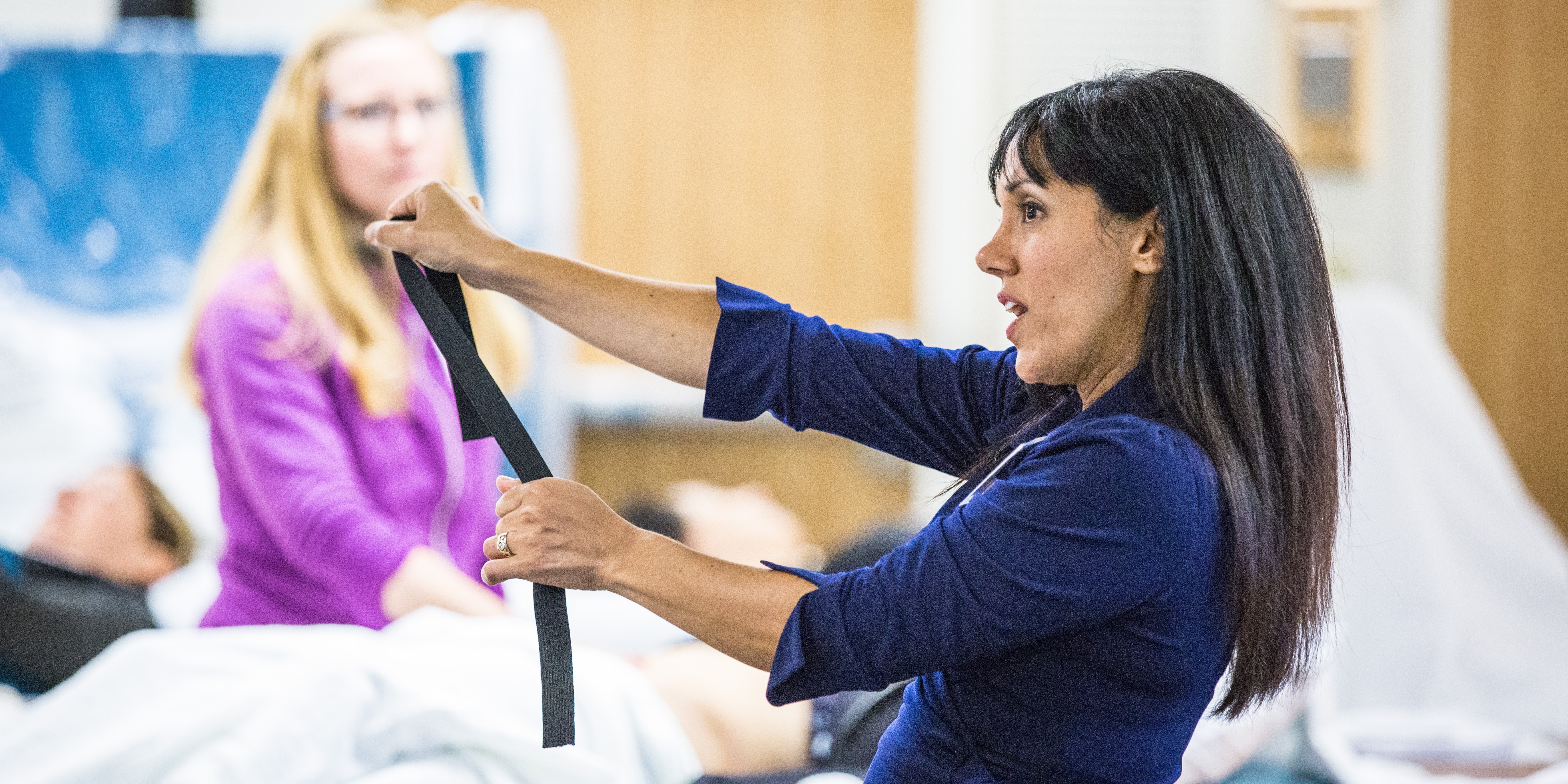
In the past decades, evidence has been established showing the importance of the local stabilizing muscles, including the transverse abdominis, the lumbar multifidus, and the pelvic floor muscles on the stability of the pelvic ring and lumbar spine. Many therapists have embraced this knowledge and incorporate a specific stabilization program into their plan of care for patients with dysfunction in the lumbar spine or pelvic ring. This plan of care can be more time consuming for the therapist since we are no longer simply prescribing strengthening exercises to the patient, but instead are using neuromuscular re-education to retrain recruitment patterns and improve motor control. For many patients, learning to activate these muscles can be very difficult and frustrating. This is where biofeedback comes in. For years, women’s health therapists have been using biofeedback to retrain the pelvic floor muscles. Biofeedback is the process of bringing unconscious physiological processes to consciousness and gaining control over it. These same principles can be used in retraining the lumbar multifidus and the transverse abdominis, in addition to the pelvic floor. Currently, biofeedback methods used in treating low back pain include rehabilitative ultrasound imaging (RUSI) and the pressure cuff. My preferred method is using RUSI. This tool not only provides biofeedback for the patient but allows for real-time assessment of the muscle activation. Thus, providing the clinician valuable feedback on timing of recruitment and strategy used by the patient that would not be assessed with palpation alone.
Giggins et al recently released a literature review that covered different forms of biofeedback used in rehabilitation. Interestingly, there is little evidence to support the use of pressure cuff. Some of the research reported found significant increases in gluteus medius and internal oblique activity. In 2013, Grooms et al also determined that correlation and likelihood coefficients indicate that the pressure cuff is likely of minimal value to detect transverse abdominis activation. I personally have had patients referred to me from other therapists to confirm whether or not they were activating their transverse abdominis. In previous treatments, the patients had been using the pressure cuff and palpation by the therapist as confirmation of proper activation. What I found was consistently the patients were not achieving good contractions in their transverse abdominis muscles. Most of these patients were able to learn in one or two sessions how to properly contract their transverse abdominis, and were later able to progress to performing a co-contraction of all the local stabilizers during motor tasks.
Hides et al published a study that demonstrated a specific stabilization rehabilitation program using RUSI was successful in decreasing pain, increasing muscle cross sectional area, and improving motor control in elite cricket players that had previously experienced low back pain. This is a great example of how neuromuscular re-education using RUSI works for the patient when the therapist is using it as both an assessment tool, as well as a biofeedback tool.
So far the limiting drawback of RUSI is the cost of the ultrasound unit. However, companies are constantly coming out with more affordable units making this useful tool more available for clinics. If you are interested in learning more about using US imaging as both an assessment and biofeedback tool join me in Seattle this May for a course that addresses the use of Rehabilitative Ultrasound Imaging in conjunction with a specific stabilization program.
References:
Giggins, Persson, Caulfield. Biofeedback in Rehabilitation. Journal of Neuroengineering and Rehabilitation.2013; 10:60.
http://www.biomedcentral.com/content/pdf/1743-0003-10-60.pdf
Grooms, Grindstaff, Croy et al. Clinimetric Analysis of Pressure Biofeedback and Transversus Abdominis Function in Individuals With Stabilization Classification Low Back Pain. JOSPT.2013; 43(3):184-193
http://www.jospt.org/doi/abs/10.2519/jospt.2013.4397
Hides, Stanton, Wilson et al. Retraining motor control of abdominal muscles among elite cricketers with low back pain. Scand J Med Sci Sports.2010; 20: 834-842.
This post was written by H&W instructor Allison Ariail PT, DPT, CLT-LANA, BCB-PMD. Allison will be instructing the Pelvic Floor Level 1 course Boston this October.
Several weeks ago some of my fellow faculty members and I were discussing the resting tone of the pelvic floor. These days we take it for granted that we know there is constant low-level activity in the pelvic floor and anal sphincter in order to provide continence. However, how did this information come about? I took it upon myself to do some research to find out the beginnings of this knowledge. What I found was interesting and thought I would share.
In the late 1940’s and early 1950’s the belief was held that the pelvic floor and external anal sphincters were inactive at rest, like other striated muscle throughout the body. Activity was believed to be initiated by afferent impulses from the rectal ampulla and anal canal. In 1953 Floyd and Walls found activity in the external anal sphincters at rest, even during sleep. In 1962 Parks, Porter, and Melzak published a study examining the pelvic floor muscles and the external anal sphincters using electromyography recordings. They also found activity in these muscles at rest. They hypothesized the activity was maintained by spinal reflex. These researchers looked at the activity in a healthy population, a paraplegic population, and a population that had undergone a rectal excision. When examining the paraplegic population (all subjects had complete SCI injuries above L3), they did identify activity of the pelvic floor at rest.
With respects to the rectal excision population, they examined patients whose rectums were removed, but the somatic muscles, external sphincters, and the levator ani remained with innervation intact and the muscles were sutured to provide a muscular pelvic floor. These patients also exhibited activity in the pelvic floor and anal sphincter at rest. These patients were important to the study in order to rule out that the reflex was not coming from somewhere in the rectal wall. Additionally, these researchers discovered this resting activity that was present the anal sphincter was inhibited during defecation in response to a certain degree of rectal distension.
So what did all of this new information mean to these researchers? It meant that the pelvic floor and external anal sphincter were unique due to the fact they were activated at rest, and without this activation continence would not be maintained. They determined the activation to be reflex and termed it “postural reflex of the pelvic floor.” Additionally, they termed the inhibition due to rectal distension “the rectal inhibitory reflex,” which also was due to a reflex arc. This new information was groundbreaking for the time and lead to other research that provided us with the knowledge that we have today! Thank goodness for these researchers as well as the many others who have furthered the advancement of knowledge about the pelvic floor!
Learn more about Allison and the Pelvic Floor Series by visiting our website!
1. Floyd, Walls. Electromyography of sphincter ani externus in man. J. Physiol. 122: 599, 1953.
2.Parks, Porter, Melzak. Experimental study of the reflex mechanism controlling the muscles of the pelvic floor. Dis. Colon Rectum. 5 (6): 407. 1962.
This post was written by H&W instructor Allison Ariail, PT, DPT, CLT-LANA, BCB-PMD. Allison will be instructing the Care of the Postpartum Patient course in Houston in June.
As Mother’s day weekend approaches, I take time to think about the dramatic changes in life that occur with the birth of a baby! No one is quite prepared for how much their life will change with the birth of their child, especially their first child! There are numerous changes that occur in a woman’s life during the pregnancy and into the postpartum time, both emotionally and physically. Any woman who has had a baby knows our bodies do not revert back to the exact body we had prior to pregnancy. New moms may be left with changes in their body that can greatly affect their function. Physical therapy in the postpartum time can greatly improve a woman’s well-being and function. We can treat a woman for back pain, diastasis rectus separations, incontinence, thoracic outlet syndrome, nerve damage that occurred during delivery, and many more issues a woman may present with. We also are a listening ear for the new mom going through many changes and hormonal upheaval. It is important to stay open and listen in a non-judgmental way. New moms are inundated with unsolicited advice in a way that no other patient population is. Having a safe place to come and get treated physically can help her emotionally as well.
During pregnancy and the postpartum time many habits are formed that if not changed can influence and shape how a woman lives the rest of her life. For example, night time voiding is common for pregnant women. If a woman continues to void every time she gets up with the baby in the middle of the night once she delivers, she may continue or even worsen her habit, thus creating an issue that will greatly affect her overall sleep health and well-being for the rest of her life. Having an objective person educate a woman about some of these habits can be very enlightening for an individual!
Receiving therapy in the postpartum time can influence a woman’s overall health in the immediate future as well as down the road. There are special things to consider when treating a postpartum woman and a women’s health therapist is the best person to treat her.
You can learn about special topics that affect a postpartum woman in Care of the Postpartum Patient course. The next time this course is being offered is June7-8 in Houston, Texas. So as mother’s day comes upon us, let’s celebrate the amazing journey we and other moms take in becoming a mom. Let us embrace the remarkable changes that occur physically and emotionally and thank our own mothers, as well as ourselves, for being willing to undergo these changes!

This post was written by H&W instructor Steve Dischiavi, MPT, DPT, ATC, COMT, CSCS. Dr. Dischiavi will be instructing the course that he wrote on "Biomechanical Assessment of the Hip and Pelvis" in Virginia this August.

In an outpatient sports medicine clinic the traditional model of physical therapy evaluation typically includes the therapist reviewing a patients chart and subjective symptom questionnaire of some sort. Then the therapist will bring the patient to an area to begin a subjective history and then onto a physical exam. After these procedures have been completed the therapist will typically assign a working clinical diagnosis and then begin treatment. In short, I would like to suggest a paradigm shift to this traditional model of thinking. Instead of starting the exam on a table with a static assessment of the structures involved and identifying the pain generator, I suggest the therapist begin with a specific set of movements used as an evaluative tool to identify movement dysfunction within the anatomical system as a whole.
All human interactions on earth occur between ground reaction force and gravity, our bodies are mostly just stuck in the middle of this constant battle and typically we succumb to whichever power exposes the weakest link in our biomechanical chains. One of the reasons the biomechanical chains in our bodies are so pliable and vulnerable to constant ground reaction force and gravity acting on them is because we are basically bones or struts suspended in a bag of skin all connected by soft tissue. Suggesting that without skin, fascia, and connective tissue supporting us, we would collapse to the ground in a pile of bones! Ingber (1997), suggested this concept, known as tensegrity, was the “architecture of life.” So in summary, the tensegrity structures are mechanically stable not because of the strength of the individual bones, but because of the way the entire human body distributes and balances mechanical stresses through the use of polyarticular muscle chains called slings. There will be more on slings in the upcoming blogs.
If there is truly a paradigm shift with the way we initially assess our clients and we begin our evaluations with whole system movement patterns it would be because we want to actually see how this tensgerity model is essentially collapsing under the stress of gravity. We would get a first hand glimpse in real time how energy leaks and blocks occur during human movement. These concepts are the foundation for the course Biomechanical Assessment of the Hip & Pelvis.
Ingber, D.E. (1997). Tensegrity: the architectural basis of cellular mechanotransduction.
Annual Review of Physiology, 59, 575-59.
Dr. Dischiavi's course is designed to elevate the participant’s skill level through advanced training in hip and pelvic biomechanics, functional “slings” created by the myofascial system, and through use of sports medicine theory and applied science. Biomechanical Assessment of The Hip & Pelvis will be taking place in August in Arlington, VA

There's a lot going on in the world of pelvic rehab, and continuing education is no exception! This March, Herman & Wallace is hosting NINE courses around the country. It's a lot to keep up with, so we thought you might appreciate a brief overview of what's coming up next!
Where's this pain coming from?
Pelvic pain can have many sources, and Elizabeth Hampton wants to help you quickly get to the source. Finding the Driver in Pelvic Pain empowers you to play detective in order to help even the most complex patients. Don't miss out on Finding the Driver in Pelvic Pain in San Diego, CA on March 4-6, 2016
What goes in eventually comes out
How important is a good diet? For most of us eating healthy is important, and for many pelvic rehab patients it is a necessity. That's why Megan Pribyl wrote her "Nutrition Perspectives for the Pelvic Rehab Therapist" course. This beginner level course is intended to expand the your knowledge of the metabolic underpinnings for local to systemically complex disorders. Don't miss out on Nutrition Perspectives for the Pelvic Rehab Therapist - Kansas City, MO - March 5-6, 2016!
There's fascia everywhere!
Fascial mobilization is a rising star in pelvic rehab treatment techniques, and Ramona Horton is excited to share it with you! "Mobilization of the Myofascial Layer: Pelvis and Lower Extremity" is the best opportunity you'll get to learn about the evaluation and treatment of myofascia for pelvic dysfunction. Check it out on our continuing education course page. Ramona will be teaching these techniques in Santa Barbara, CA on March 11-13.
Giving birth hurts
Sometimes the newborn is the one to get all the attention, but what about the new mother? Be sure that you can help postpartum women with symptoms like postural dysfunction, pelvic girdle dysfunction, diastasis recti abdominis and more by attending Care of the Postpartum Patient in Seattle, WA this March 12-13, taught by the wonderful Holly Tanner!
Vulvar pain is easy to have and hard to lose
12% of women in the US have vulvar pain for 3 or more months at some stage in their life. It takes a multidisciplinary approach to address all the causes and co-morbidities, and that is exactly what you'll get at Dee Hartmann's Vulvodynia: Assessment and Treatment in Houston, TX on March 12-13, 2016. Dee aims to address the vicious cycle of pain, visceral and sexual dysfunction, and the general hit to quality of life that patients with vulvodynia suffer from.
The challenge of SI joint pain
The sacroiliac joint, pelvic girdle, and pelvic ring sure can take a beating, and Peter Philip knows how to keep you moving. Through exercise and stabilization, the pelvic rehab practitioner can quickly treat pain in the lumbopelvic-hip complex. Learn all about the direct and indirect anatomy that influences the sacroiliac joint, and then get ready to find and treat the source of pain and dysfunction in Sacroiliac Joint Treatment in Minneapolis, MN on March 19-20, 2016.
Taxes and Menopause
The menopause transition is not something many people look forward to. For some women it goes more smoothly than others, and it's the less fortunate ones who need access to a well-trained pelvic care professional. Michelle Lyons is flying in from Ireland to help you to become that pro! Be it vaginal atrophy, sexual health dysfunction, pelvic organ prolapse, or any other of the myriad possible symptoms of menopause, you'll be equipped to handle them all after attending Menopause: A Rehabilitation Approach in Atlanta, GA on March 19-20, 2016.

The addition of the International Fascia Research Congress onto the scene of educational conferences has ignited an increased focus on understanding how fascia works in the body. Of course, we know fascia plays a role in compartmentalizing and separating various structures in the body, yet we also know that fascia must allow communication with the rest of the body. Is fascia simply a structural tissue that plays a mechanical role? Or does fascia hold memories, accessible during bodywork, as discussed in this article?
It may seem logical that fascia could contribute to compressive forces on the skeleton, on muscles and neurovascular structures, possibly contributing to musculoskeletal disease. Is myofascial tension sufficient to cause enough mechanical stress to create micro-damage and histochemical responses? Can this then lead to ankylosing spondylitis or axial spondyloarthritis, as discussed in this article published in Arthritis Research & Therapy? And if fascial thickness and tension is a proposed culprit of conditions such as compartment syndrome, why did these researchers find no correlation and in fact a negative correlation between fascial stiffness in patients with compartment syndrome?
Do we really know the implications of fascially-directed assessments and interventions at this time? Is the research on fascial therapy being interpreted correctly if science is still trying to figure out what fascia is, how fascia works, how fascial forces affect the body and body functions? If we don't yet understand the intricacies of the neurophysiological mechanisms that drive fascia, should we jump to conclusions about the science that may or may not be measuring the right variables? (To this end, is a test of the fascial strength meaningful if taken from a biopsy now that the tissue is disconnected from the nervous system?)
I am not a fascial researcher, and I appreciate those who do give their time and energy towards working on these questions. As a pelvic rehabilitation provider, I know that fascial relationships within the pelvis are multi-faceted and somewhat unique: the obturator internus (OI) attaches directly into a thick fascial line running between the OI and the levator ani muscles. The potential implications of this relationship on muscle strength and tension are constant clinical considerations, and ones that we hopefully will know more about as tools such as functional MRI lend improved data.
Clinicians who utilize myofascial assessments and treatment have more understanding of the role of substances such as hyaluronic acid in fascial health, yet we are still searching for accurate ways to describe how stretching and connective tissue manipulation can ease chronic pain. While we continue to explore the science behind the techniques, you can further your knowledge of fascia and fascial techniques at the continuing education course Myofascial Release for Pelvic Dysfunction, offered for the last time this year in Ohio this month.
Does wearing a pelvic belt affect the activation of the gluteus maximus and gluteus medius muscles in healthy males? Recent research asked this question, and the results, although difficult to extrapolate to other patient populations, are interesting. Surface electromyography (sEMG) amplitude was measured in 20 male patients during 6 exercises, and the amplitude during the exercise was compared to a maximum voluntary contraction. The findings demonstrated that muscle activation increased in the gluteus maximus when a pelvic belt was worn. Activation in the gluteus medius was unchanged for all exercise except during the clam exercise when the gluteus medius was noted to be more active.
Mean age in the study was 23 years, and all participants reported a lack of disease or injury. All were able to complete the exercises without pain. The 6 exercises that were instructed by an experienced physical therapist included hip clam, side lying hip abduction, single limb squat, single limb deadlift, frontal planar lunge, and frontal planar hop. Each exercise was performed 3 times, the order of exercise was randomized, and the dominant limb was used.
The authors bring up interesting points and hypotheses in relation to the sEMG findings. In a patient who presents with lumbar pain and delayed gluteus maximus activation, can a pelvic belt be utilized to improve muscle activation and therefore pelvic stability? Is adding a belt such as the COMPRESSOR belt used in this study valuable for allowing a patient to optimally complete dynamic activities, or does the belt inhibit gluteus medius activity by providing support that the muscles are supposed to provide? Most research invites us to consider the clinical implications of an intervention or a strategy, and the rehabilitation provider must assess the value of the strategy for that particular patient.
For practitioners who are interested in fine-tuning skills in lumbopelvic and hip assessment, Tracy Spitznagle, instructor in the Physical Therapy program at Washington University, will teach the Movement System Approach to Musculoskeletal Pelvic Pain: Lumbar, Hip, and SI Joint in April in Houston, TX. In this 2-day continuing education course, participants will learn to recognize movement impairment syndromes, perform movement tests, and develop a corrective exercise program based on a specific movement examination.
All Upcoming Continuing Education Courses

Mobilization of Gastrointestinal Visceral Fascia Satellite Lab Course - Self-Hosted - December 6 - 8 2024
Dec 6 2024 - Dec 8 2024

Mobilization of Visceral Fascia: The Gastrointestinal System - Phoenix AZ - December 6 - 8 2024
Dec 6 2024 - Dec 8 2024

Mobilization of Visceral Fascia: The Gastrointestinal System Satellite Lab Course - Woodbury NY - December 6 - 8 2024 - SOLD OUT
Dec 6 2024 - Dec 8 2024

Mobilization of Visceral Fascia - Gastrointestinal Satellite Lab Course - Raleigh NC - December 6 - 8 2024
Dec 6 2024 - Dec 8 2024

Mobilization of Visceral Fascia - Gastrointestinal Satellite Lab Course - Medford OR - December 6 - 8 2024
Dec 6 2024 - Dec 8 2024

Mobilization of Visceral Fascia: Gastrointestinal Satellite Lab Course - Milwaukee WI - December 6 - 8 2024
Dec 6 2024 - Dec 8 2024

Mobilization of Visceral Fascia - Gastrointestinal Satellite Lab Course - Ann Arbor MI - December 6 - 8 2024
Dec 6 2024 - Dec 8 2024


Nutrition Perspectives for the Pelvic Rehab Therapist - Remote Course - December 7 - 8 2024
Dec 7 2024 - Dec 8 2024

Biofeedback for Pelvic Muscle Dysfunction Satellite Lab Course - Self-Hosted - December 7 2024
Dec 7 2024

Sacral Nerve Manual Assessment and Treatment - Remote Course - December 7 - 8 2024
Dec 7 2024 - Dec 8 2024


Pelvic Function Level 2B - Satellite - Colorado Springs CO - December 7 - 8 2024
Dec 7 2024 - Dec 8 2024


Pelvic Function Level 1 - Satellite - Denver CO - December 14 - 15 2024 - SOLD OUT
Dec 14 2024 - Dec 15 2024
Pelvic Function Level 1 - Satellite Lab Course - Lone Tree CO - December 14 - 15 2024 - SOLD OUT
Dec 14 2024 - Dec 15 2024

Pelvic Function Level 1 - Satellite - Jacksonville FL - December 14 - 15 2024
Dec 14 2024 - Dec 15 2024

Pelvic Function Level 1 - In-Person - New York NY - December 22 - 23 2024 - SOLD OUT
Dec 22 2024 - Dec 23 2024


Pelvic Function Level 1 - In-Person - Philadelphia PA - January 11 - 12 2025 - SOLD OUT
Jan 11 2025 - Jan 12 2025

Oncology of the Pelvic Floor Level 1 - Remote Course - January 11 - 12 2025
Jan 11 2025 - Jan 12 2025

Modalities and Pelvic Function - In-Person - Sacramento CA - January 11 - 12 2025
Jan 11 2025 - Jan 12 2025

Pediatrics Level 1 - Treatment of Bowel and Bladder Disorders - Remote Course - January 11 - 12 2025
Jan 11 2025 - Jan 12 2025


Pelvic Function Level 1 - Satellite - Knoxville TN - January 18 - 19 2025 - SOLD OUT
Jan 18 2025 - Jan 19 2025


Pelvic Function Level 1 - Satellite - Manchester IA - January 18 - 19 2025
Jan 18 2025 - Jan 19 2025

Pelvic Function Level 1 - Satellite - Long Beach CA - January 18 - 19 2025
Jan 18 2025 - Jan 19 2025


Pelvic Function Level 1 - Satellite - Fort Lauderdale FL - January 18 - 19 2025
Jan 18 2025 - Jan 19 2025

Pelvic Function Level 2B - Satellite - Bradenton FL - January 18 - 19 2025
Jan 18 2025 - Jan 19 2025
Pelvic Function Level 2B - Satellite - Commerce Township MI - January 18 - 19 2025
Jan 18 2025 - Jan 19 2025