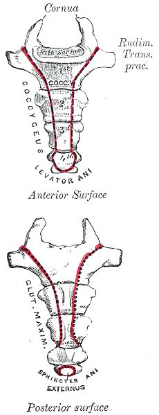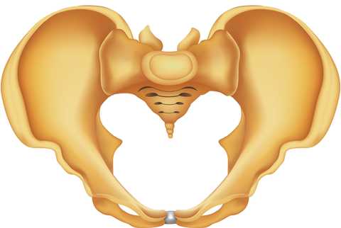
A recent publication of a case series proposes that clinicians use intranasal calcitonin, a medication prescribed for acute vertebral fractures in osteoporotic patients, for acute coccyx pain. This medication is administered as a nasal spray, has been demonstrated to have analgesic properties, and is theorized to have effects of beta-endorphin production, and inhibition of prostaglandin and cytokine production. Nasal calcitonin, a synthetic drug, is reported to have fewer and mild side effects when compared with the injectable form of the medication.
Of the 8 subjects treated and described in the case series, all had a fall on the tailbone, either landing hard into a chair on onto the stairs or floor. Some were seen within 3 months of injury date, and others were treated after 3 months from injury. The authors report that in the subjects who were treated acutely, 3 of 5 had at least 50% improvement in pain, without injections or surgery. The subjects treated in a chronic pain condition had similar levels of improvement but with the addition of coccyx injections for treatment.
Based on the lack of a control group, the small number of subjects treated, and because the mechanism of nasal calcitonin is unknown as far as effects on fracture healing in the coccyx, we would not, as rehab professionals, look to our referring providers to add nasal calcitonin routinely in the management of acute coccyx fracture. This article may, however, lead to interesting conversations with referring providers who are in a position to consider the addition of calcitonin for the proposed analgesic effect and potential bone healing augmentation. (The authors do suggest using the smallest effective dose for the shortest duration required.)
Clinically, is the difference in recovery for acute versus chronic coccyx pain of interest? Ideally, we would get to work with patients very soon after an injury, so that education is in place about self-care, self-treatment, optimal movement and thought patterns. The earlier that we can help a patient alleviate muscle tension and pain, and increase movement within tolerance, the better the outcomes usually are for the patient, as patterns of inactivity, fear, and muscle guarding are less likely to have set in. To learn much more about treatment of the coccyx, join faculty member Lila Abate in New Hampshire this September for the Coccyx Pain: Evaluation and Treatment continuing education course.
This post was written by H&W instructor Dawn Sandalcidi, PT, RCMT, BCB-PMD. Dawn's course that she wrote on "Pediatric Incontinence" will be presented in in South Caroline this August.

Years ago when my oldest daughter was 4 years old and in Pre-school I received an urgent call at the office that she had an accident. Immediately my head began to race, “What hospital is she in?” “What did she break?” Then the director informed me she wet her pants. I collapsed in my chair with a huge sense of relief and I began to ponder “Did she have an ‘accident’ or did her bladder leak?
Merriam-Webster defines an accident as:
- 1. An unforeseen and unplanned event or circumstance
- 2.An unexpected and medically important bodily event especially when injurious
- 3.Used euphemistically to refer to an involuntary act or instance of urination or defecation
Now lets think about that. How would you feel if someone approached you after noticing a smell or a wet spot and asked you “Did you have an accident?” My first thoughts are maybe shame, embarrassment, guilt or failure. “I” had an accident. Children feel without easily being able to express these emotions thus internalizing their feelings. This then can be expressed with inappropriate behaviors.
When I work with children, and adults for that matter, I frame the conversion with the physiology of the anatomical structure that is unable to do the job it is designed to do. I teach the children about their anatomy and bladder/bowel function and I am clear to let them know that their bladder and/or bowel had a leak, they did not. It takes ownership away from the person and places it on the body part that is currently dysfunctional. At that point we discuss we can re-train the body part to do the job they were designed to do. The kids become empowered that they will be able to become “The Bladder/Bowel Boss”.
To learn more about Dawn's course visit Pediatric Incontinence
Earlier this year, when I came across this journal article about developing an interdisciplinary clinic for patients with endometriosis and pelvic pain, I was pleased to see that rehabilitation is included in this team setting. Upon reaching out to Susannah Britnell, physiotherapist, she agreed to answer some questions about her clinic and about her passion for pelvic rehab. Below you can read her responses to my questions. Stay tuned for Part 2 that will be published next week!

How long have you been working in the interdisciplinary clinic?
I’ve been working at the BC Women’s Centre for Pelvic Pain and Endometriosis in Vancouver, BC, since October 2012. We are a provincially funded program for women with chronic pelvic pain from all over the province of BC, Canada.
How long have you practiced?
I have been practicing since 1997 and love it! It is amazing how our practice can grow and move into different areas. We are truly lucky we have these opportunities.
What other settings have you worked in?
I have worked in hospital and private practice settings. I found my grounding in musculoskeletal physiotherapy, obtained my manual and manipulative therapy diploma in 2001, and worked in obstetrics for 10 years at BC Women’s hospital. I then started incorporating pelvic floor physiotherapy into my practice and developed a strong interest in chronic pain and the biopsychosocial approach to pain management. In addition to clinical practice, I teach the obstetrics component at UBC School of Rehabilitation as an annual guest lecturer and am an instructor for physiotherapy Rost Therapy courses for pelvic girdle pain, which I often co-instruct with Cecile Röst, a Dutch Physiotherapist.
How did you get involved with the interdisciplinary group?
Were you involved in the development of the center?
Our gynecologists, Dr Allaire and Dr Williams have been working at the Women’s Hospital and Health Centre for many years and identified the need for an interdisciplinary program for women with pelvic pain. After getting the funding in place, they hired a counsellor, physiotherapist, and nurse to be a part of the team. We now also have another gynaecologist, Dr Yong, as well as a Fellow. The gynecologists, counsellor, nurse, and myself along with administrative program manager worked together to develop the program. We have also had input from a Pain Specialist/anesthesiologist. I am so grateful and thankful to be a part of such a supportive and skilled team. As a physiotherapist, it truly is wonderful to have such support from the physicians
Our program consists of a full day workshop which includes pain education, life style changes, mindfulness based stress reduction and meditation techniques. We address fear of movement and give strategies for pacing and grading activity and we also include stretches, posture, and positioning advice. Women then book individual physiotherapy and counselling appointments so we can work on more specific concerns. Physiotherapy treatment may include some passive treatment techniques, but only as a bridge to more active self management. Patients will always have an active plan from the first visit. It is important that we don’t reinforce a sense that the woman needs someone to “fix” her concern and that she has no ability or power to make changes with her pain.
Our nurse is in contact with the patients from the beginning and is available if they have any questions as they go through the process. To learn more about the program, you can go to www.womenspelvicpainendo.com or contact Susannah at sbritnell@cw.bc.ca.
To discover more information about working with chronic female pelvic pain, you can attend one of our series courses such as Pelvic Floor Level 2B, or our advanced course, Pelvic Floor Level 3
This blog was written by Carolyn McManus, PT, MS, MA, who will be presenting at the APTA's Virtual NEXT conference in North Carolina. Carolyn is instructing a new course with Mindfulness-Based Biopsychosocial Approach to Chronic Pain. This course will be offered November 15-16, 2014 in Seattle, WA.

We all know a highly stressed patient will have a more complicated and prolonged healing process than a non-stressed patient. Recent research is finally illuminating possible mechanisms causing the amplification of pain by stress. For example, stress has been shown to enhance muscle nociceptor activity in rats. (1) In this study, water avoidance stress produced mechanical hyperalgesia in skeletal muscle and a significant decrease in the mechanical threshold of muscle nocicpetors, a nearly two fold increase in the number of action potentials produced by a fixed intensity stimulus and an increase in conduction velocity from 1.25 m/s to 2.09 m/s! Researchers suggest these effects are due, at least in part, to catecholamines and glucocorticoids acting on adrenergic and glucocorticoid receptors on sensory neurons.
I always talk about this study with my patients who have persistent pain. It helps them understand that pain can escalate, not because of tissue damage, but because of the effects of stress hormones on their nerves. They understand that if they persist in escalating their stress reaction they will limit their healing potential. There is more research on this topic and strategies to help patients self-regulate their stress reaction that I look forward to discussing in my upcoming course in November.
Learn more about Carolyn's course Mindfulness-Based Biopsychosocial Approach to Chronic Pain
In Turkey, the rate of daytime urinary incontinence (DUI) was studied in primary school-aged children. A questionnaire was completed by parents of 2164 students. The Dysfunctional Voiding and Incontinence Symptoms Score Questionnaire was utilized and includes 14 questions about daytime and nighttime symptoms, voiding and bowel habits, and quality of life.
The population studied included approximately half boys and girls, with nearly half living in rural versus urban settings, and of a mean age of 10 years old. The overall prevalence of DUI was 8% (8.8% in girls, 7.3% in boys), and decreased with increasing age in this study population of children in 1st through 8th grades. 57.8% of those who did experience involuntary loss of urine were wetting less than 1x/day, 26.6% were wetting 1-2 times/day, and 15.6% were wetting greater than 2x/day. Urge incontinence was reported in nearly 59% of the children with DUI.
Independent risk factors for DUI included age, maternal educational level, family history of daytime wetting, urban versus rural setting, history of constipation or urinary tract infection (UTI), and urinary urgency. The authors conclude that "…educational programs and larger school-based screening should be carried out, especially in regions with low socioeconomic status." In this study, one of the strategies used to increase the survey response rate was to send medical and public health residents to the schools to speak about the study on 2 occasions.
Despite the numbers of therapists who have previously trained with educators such as faculty member Dawn Sandalcidi in her coursework for pediatric bowel and bladder function, the number of therapists focusing on pediatric pelvic health remains small while the need is great. Therapists who wish to expand community reach to pediatric urologists and pediatricians, and to serve the children who may unfortunately become adults who have bowel and bladder dysfunction, have the opportunity to attend the Pediatric Incontinence and Pelvic Floor Dysfunction continuing education course in Greenville, South Carolina this August.
In women with pelvic pain, is a pudendal nerve block effective, and how does the effectiveness correlate with findings of the history and clinical exam? These were the questions posed by a study published in 2012. Sixty-six patients were given a standardized pudendal nerve block, with Visual Analog Scores (VAS) and presence of numbness recorded prior to and up to 64 hours after the block. Inclusion criteria for the study involved having spontaneous or provoked pain in the distribution of the pudendal nerve, and patients were excluded if significant psychosocial issues, neurogenic or neuromuscular disorders, contraindication to sedation or allergy to utilized medication was present. A detailed history and physical examination was completed.
The pudendal nerve block was administered transvaginally and digitally, under sedation, in a lithotomy position. Following data collection, the researchers found that the presence of a positive Tinel's sign (palpation medial to the ischial spine for assessment of pain reproduction), a prior history of vulvovaginal candidiasis, or symptom worsening in the sitting position was associated with a return of the pain prior to the numbness wearing off. 92.4% of the subjects reported a "positive" response to the block, with varied lengths of time of symptom reduction. Nearly 87% of the subjects reported a reduction in one or more symptoms. This study only studied subjects for 64 hours, therefore it is not possible to discuss from this research the long-term implications of a pudendal nerve block in women with pudendal neuralgia. The authors did find a correlation between prior traumatic events including birth injuries, herniated discs, and fractures of the coccyx, pelvis or sacrum.
What does this research tell us about the role of pudendal blocks in the assessment and treatment of female pelvic pain? As already mentioned, the brevity of the data collection (only up to 64 hours) in addition to application of a non-guided block limit the ability to extrapolate this information to any long-term results. However, the correlations to clinical history and the return to pain prior to numbness ending may provide useful information as further clinical research is completed. The numbness was found to have inconsistent effects on a patient's symptoms such as bladder, bowel, sexual dysfunction, or sitting, and further research could measure the effects of a block on these functions. If you would like to discuss pudendal blocks with experts on pudendal dysfunction, sign up for the remaining spots in our August San Diego course!
Learn more about the Pudendal Neuralgia Assessment and Treatment course that we are holding at Comprehensive Therapy Services later this year.
This post was written by H&W instructor Dawn Sandalcidi, PT, RCMT, BCB-PMD. Dawn's course that she wrote on "Pediatric Incontinence" will be presented in in South Caroline this August.

I will never forget the morning I was called by one of my referring pediatricians to tell me an 11-year-old boy with fecal incontinence hung himself because his siblings ridiculed him. If you ever ask me why I do what I do, I will tell you so that nothing like that would ever happen again.
When we think of pediatric bowel and bladder issues we primarily focus on the physiologic issue itself and treating the underlying pathology. I think it is imperative to teach a child that she/he did not have a leak but their bladder or bowel had a leak. It makes the incident a physiological problem and not a problem of the child.
It is not always apparent how much the child is suffering from issues with self-esteem, embarrassment, internalizing behaviors, externalizing behaviors or oppositional defiant disorders. Dr. Hinman recognized theses issues years ago (1986) and commented that voiding dysfunctions might cause psychological disturbances rather than the reverse being true. Dr. Rushton in 1995 wrote that although a high number of children with enuresis are maladjusted and exhibit measurable behavioral symptoms, only a small percentage have significant underlying psychopathology. In more recent studies by Sureshkumar, 2009; Joinson 2007 it was noted that elevated psychological test scores returned to normal after the urologic problem was cured. Lettgen et al. 2002, Kuhn et al, 2008, van Gontard, 2012 all reported that children with urge incontinence are distressed by their symptoms but the family functioning is intact.
I frequently get testimonials from my patients. I would say the common denominator is the child and/or parental report that the child is “much better adjusted,” “happier”, “come out of his shell”, “more outgoing”, “making friends.” As a side note -- they’re happy they don’t leak anymore.
The International Children’s Continence Society (i-c-c-s.org) is filled with standardization documents that support the work we do to take care of kids with elimination issues. The work we do to take care of these kiddos in not only necessary but also mandatory to avoid these psychological disorders.
To learn more about Dawn's course visit Pediatric Incontinence
Read more about what Dawn does in PT in Motion
A systematic review from July of 2013 addressed how interpersonal factors modulate pain. Four primary findings of the research that are found to affect pain-related responses are as follows: the degree to which social partners were active (or perceived to be able to be active), the degree to which participants could perceive the specific intentions of the social partners, the pre-existing relationship between the subject and the social partner, and individual differences in relating to others (including coping styles).
Some patients are negatively impacted by a partner who is attentive to pain, with increased reports of pain, worse pain outcomes when together versus alone, and with longer lasting pain states. On the other hand, some patients respond favorably to a partner's support, with lower reports of pain states when offered support such as holding the hand of a partner. Because each person's relationship and response to the pain within the context of the relationship varies, the impact of a partner on perceived pain is also varied. The authors, after describing details and evidence of pain modulation research, conclude the following: "Specifically, interpersonal exchanges affect precision or salience by socially signaling the safety or threat of the impending stimulus itself or the environment in which the stimulus occurs."
How can we take this information to heart within the pelvic rehabilitation practice? One of the ways that we offer support to a couple is by inviting a partner to attend a clinic session where he or she can learn to assist in application of soft tissue release techniques. The partner has the opportunity to be validated in both the gratitude that the therapist offers to the patient and partner for attending, and also in the fact that pelvic pain is commonly encountered. Because pelvic pain can interfere with a couple's intimacy, having such validation about the physicality of pain, when present, may be useful in a relationship. When a partner learns how to be of help in the healing process, this may also affect the factors mentioned in the cited research article, specifically, how partners are perceived to be able to be active or perception of specific intention.
It has been my clinical experience that partners are very specific in intention once being trained in how to help with pelvic pain, and that intention is to be a part of the healing process. It has also been an observation that if a partnership is struggling with their relationship, that issues can surface once asked to engage in pelvic muscle rehabilitation. This might mean that a patient chooses to pause rehabilitation and enter psychological counseling or other healing work. It also might mean that the patient chooses to not request help of the partner in the clinic or at home for the time being.
In relation to the pre-existing interpersonal factors, we do not necessarily know the extent to which the patient, partner, or the relationship has the ability to cope with challenges, or what type of relationship is in place. For that reason, we must remain nonjudgmental, and recognize that as rehabilitation professionals, we are limited by the scope of our practice and may serve the patient best by coordinating a referral to a counselor or psychologist. If you are interested in learning more about the research as well as the practical clinical implications of pain neurobiology, we still have a few seats left in our continuing education course titled Meditation and Pain Neuroscience. This course takes place in September in Illinois, and features an accomplished physical therapist as well as a psychiatrist. This course will offer an amazing learning experience as it combines the perspectives of medicine and rehabilitation, we hope you can join us!
This post was written by H&W instructor Ginger Garner, MPT, ATC, PYT. Ginger will be instructing the course that she wrote on "Yoga as Medicine for Labor and Delivery and Postpartum" in Washington this August.

Physical therapists often see women during pregnancy and postpartum, but what can physical therapists do to foster better birth outcomes?
A 2012 study conducted in Norway underscores the importance of childbirth education, which can take place as part of patient education and counseling in physical therapy. The study looked at 2206 women with intended vaginal delivery in order to assess the association between fear of childbirth and duration of labor. Labor duration was found to be significantly longer in women with fear of childbirth, with the rate of epidural analgesia, induction, and instrumental vaginal delivery also being higher in fearful women. The authors posit that “anxiety and fear may increase plasma concentrations of catecholamines, and high concentrations of catecholamines have been associated with both enervated uterine contractility and a prolonged second stage of labour.” (Adams et al 2012).
Yoga is a mind-body intervention that is supported to lower pain perception, anxiety, reported stress, and discomfort, all variables that can improve overall birth outcomes and reduce fear of childbirth. Integrative physical therapy practice uses a biopsychosocial model, one that uses energetic, emotional, physical, intellectual, and spiritual support methods to prepare a mother for her labor, delivery, and beyond. Mason et al (2013), in a study that compared standard diaphragmatic breathing to yogic breathing, found yogic breathing to be superior in all measured areas, including increasing/affecting: 1) cardiac-vagal baroreflex sensitivity, 2) oxygen saturation, 3) oxygen absorption, 4) tidal volume, 5) vagal stimulation, 6) parasympathetic activation, and 7) overall reported physical and mental health.
Yoga can address more than just flexibility or relaxation for laboring moms. It fosters calm awareness, mind-body concentration and focus, develops postural control and lumbopelvic health, including neural and myofascial health and motor patterning, all of which, combined with conventional physical therapy practice, can be more efficacious than exercise prescription or childbirth education alone. Ginger’s course, Yoga as Medicine for Labor, Delivery, and Postpartum addresses the systems-based changes of the pregnant patient, and prepares the physical therapist to meet the needs of the laboring or new mom with a mind-body holistic perspective.
To learn more about Ginger’s course, visit Yoga as Medicine for Labor, Delivery, and Postpartum

When assessing sacroiliac joint (SIJ) stability and function, pelvic rehabilitation providers often use the single leg stand test. During transition from double-leg stance to single-leg stance, dynamic stability is required and there are several ways to describe the observed behaviors in patients. If a patient, for example, loses her balance when transitioning to a single leg stand, what is contributing to her loss of stability? Could it be the SIJ, abdominal and trunk strength, ankle proprioception, visual or vestibular deficits, or hip abductor weakness? Of course, these are not the only potential confounders to postural stability, yet represent some of the considerations of the rehabilitation therapist.
A recent study aimed to further define techniques to measure "time to stabilization" in a double-limb to single-limb stance. The authors measured 15 healthy control subjects and 15 subjects who presented with chronic ankle instability (CAI) and the researchers tested the ability to achieve stability in single-leg stance during eyes open and eyes closed tasks as well as with varied speeds of movement.
The research found that in subjects who had CAI, following transition to single-leg stance, increased postural sway was noted. In the same subjects, when performing the task with eyes closed, those with a history of ankle instability performed the transition much more slowly than those in the control group when allowed to choose their speed of transition. Prior research (described in the linked article) had postulated that time to stabilization (TTS) following double-limb to single-limb transition was an important variable to measure, yet this research did not find that the TTS was significantly different between the groups studied.
This research highlights the value of considering the total limb and trunk stability during testing of functional tasks, as well as considering visual assist versus eyes closed variables for task completion. The speed of task performance should also vary so that deficits can be perceived by the examiner. The authors of this research propose further specifications to future research that will add to the meaningfulness and accuracy of the described testing methods. They also conclude that in patients with chronic ankle instability, an altered and poorer strategy is used in the overcoming of a postural perturbation. If you would like to further explore the sacroiliac joint examination, evaluation and treatment, and discuss concepts of stability, join Institute faculty member Peter Philip at the continuing education course Sacroiliac Joint Treatment this July in Baltimore!
By accepting you will be accessing a service provided by a third-party external to https://hermanwallace.com/


