Pelvic Rehab Report
The official Herman & Wallace blog. New blogs post every Friday on topics relating to the field of pelvic floor dysfunction.

Rachna Mehta, PT, DPT, CIMT, OCS, PRPC is the author and instructor of the new Acupressure for Pelvic Health course. Rachna brings a wealth of experience to her physical therapy practice and has a personal interest in various eastern holistic healing traditions. Her course Acupressure for Optimal Pelvic Health brings a unique evidence-based approach and explores complementary medicine as a powerful tool for holistic management of the individual as a whole focusing on the physical, emotional, and energy body.
Constipation is a common functional gastrointestinal disorder, with prevalence in the general population of approximately 20%. In the elderly population, the incidence of constipation is higher compared to the younger population, with elderly females suffering more often from severe constipation (1).
Is there a magic button in the perineum that makes it easier to defecate? In case you have wondered, the answer is YES!!!
A study done recently by Dr. Ryan Abbott and colleagues at UCLA’s Department of Medicine found just that. A randomized control trial was conducted with 100 subjects who had functional constipation, half randomized to treatment and half to the control group. The treatment group received training in self perineal acupressure along with standard treatment options. The control group only received information about standard constipation treatment options.
Perineal self-acupressure technique was found to be remarkably effective with statistically significant and clinically meaningful improvements in Patient Assessments of Constipation Quality of Life (PAC-QOL All), modified Bowel Function Index (BFI), and the Short-Form Health Survey (SF-12v2). Patients in the treatment group also reported substantial satisfaction with perineal self-acupressure technique:
- 72% of the treatment group reported that the perineal pressure technique helped them to “break up, soften, or pass stools."
- 54% reported that the technique helped them to “avoid having hemorrhoid or lessened the impact of existing hemorrhoid."
- 72% reported that the technique helped them to “avoid or better manage the effects of constipation."
- 82% of the treatment group patients indicated that they would continue to use the technique, and 72% indicated that they would recommend the technique to family and friends (2).
In this study, perineal acupressure was applied at the Acupressure point Huiyin or CV 1 located at the perineum. Huiyin is used in Traditional Chinese Medicine (TCM) not only to treat constipation, but also a variety of conditions including impotence, hemorrhoids, rectal prolapse, and dysmenorrhea. In addition, there are several key Acu-points like St 36 on the Stomach meridian and CV 6 which can help with constipation and digestive disorders.
Acupressure is based on Traditional Chinese meridian theory in which acupuncture points are pressed to stimulate the flow of energy or Qi and these points reflect disorders of visceral conditions and organs.
Acupuncture meridians are believed to form a network throughout the body, connecting peripheral tissues to each other and to the central viscera. This tissue network is also continuous with more specialized connective tissues such as periosteum, perimysium, perineurium, pleura, peritoneum, and meninges (3).
Dr. Abbott’s study suggests that clinicians should consider incorporating perineal self-acupressure technique as a first-line treatment for constipation, along with conventional interventions such as increased exercise and dietary fiber intake. Benefits include being non-invasive and non-pharmacological treatment intervention for constipation with likely a lower risk for side effects and complications than commonly used medications such as stool softeners, fiber supplements, stimulants, laxatives, and lubricants (2).
As medical providers, we are uniquely trained to combine our orthopedic skills with mindfulness-based holistic interventions to empower our patients by giving them the tools and self-care regimens to live healthier pain-free lives.
The upcoming remote course Acupressure for Optimal Pelvic Health, scheduled for July 24-25, 2021, brings a unique evidence-based approach on the use of potent Acupressure points for treating a wide variety of pelvic health conditions including chronic pelvic pain, dysmenorrhea, constipation, digestive disturbances and urinary dysfunctions to name a few.
The course also offers an introduction to Yin yoga and explores Yin poses within each meridian to channelize energy through neurodynamic pathways with powerful integrative applications across multiple systems.
References
- Vazquez Roque M, Bouras EP. Epidemiology and management of chronic constipation in elderly patients. Clin Interv Aging. 2015;10:919-930.
- Abbott R, Ayres I, Hui E, Hui KK. Effect of perineal self-acupressure on constipation: a randomized controlled trial. J Gen Intern Med. 2015;30(4):434-439.
- Kaptchuk TJ. 2000. The web that has no weaver. Understanding Chinese medicine. Chicago: Contemporary Publishing Group, Inc.
5. Lee EJ, Frazier SK. The efficacy of acupressure for symptom management: a systematic review. J Pain Symptom Manage. 2011;42(4):589-603.

Erica Vitek, MOT, OTR, BCB-PMD, PRPC has attended extensive post-graduate rehabilitation education in the area of Parkinson disease and exercise. She is certified in LSVT (Lee Silverman Voice Treatment) BIG and is a trained PWR! (Parkinson Wellness Recovery) provider, both focusing on intensive, amplitude, and neuroplasticity-based exercise programs for people with Parkinson disease. Erica has taken a special interest in the unique pelvic floor, bladder, bowel, and sexual health issues experienced by individuals diagnosed with Parkinson disease. You can learn more about this topic in Erica's course, Parkinson Disease and Pelvic Rehabilitation, scheduled for July 23-24, 2021.
Parkinson disease (PD) non-motor symptoms can be even more impactful on quality of life than the cardinal motor symptoms most are familiar with, bradykinesia, rigidity, tremor, and postural instability. The list of non-motor symptoms is extensive affecting many body systems including cognitive, sensory, and autonomic.
Constipation is one of the most common autonomic non-motor symptoms experienced by people with Parkinson disease with studies showing 20-89% prevalence (1). As the disease progresses, individuals are more likely to experience symptoms that suggest a strong relationship between neurodegeneration and bowel dysfunction, such as, decreased frequency of bowel movements, difficulty expelling stool, and diarrhea (2). Constipation has also been hypothesized to be an early indicator for the development of Parkinson disease, and there is ongoing research in this area. It has yet to be shown that constipation is specific enough to predict the development of PD.
Developing an understanding of Parkinson disease constipation and how it differs from other individuals with constipation can have a strong impact on our recommended pelvic rehabilitation plans of care. In a study published by Zhang, M. et al., 2021, they looked at the characteristics of Parkinson disease with constipation (PDC) compared to functional constipation (FC). Functional constipation is generally defined as difficult, infrequent, or incomplete defecations (3). One of the main findings in this study was a significant difference between the groups when looking at resting rectal and anal canal pressures. In the PDC group, resting rectal and anal pressures were significantly lower. These resting pressures are mainly controlled by the internal anal sphincter resting tension which is supported by the autonomic nervous system. This leads the researchers to speculate there may be autonomic nervous system neuropathy in people with PD
They then looked at simulated defecation, which also showed that the PDC group had significantly lower rectal defecation pressure and a lower anal relaxation rate. Since rectal pressures during defecation assist in effective anal relaxation, the researchers state, “a coordinated movement disorder results” and that people with PD may have “pelvic floor cooperative motion disorder” (1). Additionally, the researchers noted that abnormal abdominal pressure is another main contributing factor to the low rectal defecatory pressure in PDC. Abdominal pressure is a key factor in driving complete and efficient rectal defecation. This is also a finding in numerous other studies in the literature unique in PDC.
The results of Zhang, M. et al., 2021 reveal the need for pelvic health practitioners to help train coordinated defecation efforts in people with Parkinson disease. In my course, Parkinson disease and Pelvic Rehab, we will have an in-depth discussion about how training defecatory dynamics is different in people with PD. Muscle training principles in this population are very unique. Understanding the underlying causal factors of dysfunction will have a significant impact when helping patients with Parkinson disease manage constipation.
- Zhang, M., Yang, S., Li, X. C., Zhu, H. M., Peng, D., Li, B. Y., ... & Tian, C. (2021). Study on the characteristics of intestinal motility of constipation in patients with Parkinson's disease. World Journal of Gastroenterology, 27(11), 1055.
- Sakakibara, R. (2021). Gastrointestinal dysfunction in movement disorders. Neurological Sciences, 1-11.
- Lacy, B. E., Mearin, F., Chang, L., Chey, W. D., Lembo, A. J., Simren, M., & Spiller, R. (2016). Bowel disorders.Gastroenterology 150 (6), 1393-1407.

This blog contains an interview with Alyson N Lowrey, PT, DPT, OCS. Alyson treats the pelvic floor patient population through an orthopedic approach, working closely with pelvic floor specialists. Alyson’s clinical interests include evaluation/treatment of chronic pain, lumbar and cervical spine disorders, foot and ankle disorders, pelvic pain, and clinical instruction. Alyson is the co-instructor for the new H&W course, Pain Science for the Chronic Pelvic Pain Population - Remote Course scheduled for July 17-18, 2021.
Q: Who are you? Describe your clinical practice.
A: I work in an outpatient hospital-based clinic where I am able to provide true 1:1 care to patients of all ages and orthopedic conditions. Since pelvic floor therapy came to our clinic, I have developed strong clinical and personal relationships with pelvic floor therapists. We have been able to successfully combine our respective expertise into a wholistic approach for improving patient’s functional outcomes. My knowledge and relationships with pelvic floor therapy have allowed me as an ortho clinician to recognize when a patient’s dysfunction may have a pelvic floor component and refer appropriately. I am also in a unique opportunity where my pelvic floor colleagues will co-treat or transition care of a patient to me to continue to improve their overall function by providing functional strengthening and neuromuscular re-education to the pelvic floor musculature and other supportive muscular systems. This relationship also allows us to treat comorbid orthopedic conditions and pelvic dysfunctions such as low back pain or SIJ dysfunction as well.
Q: How did you get involved in the pelvic rehabilitation field?
A: I became involved with pelvic rehabilitation through working in a clinic with Tara Sullivan. Her knowledge is immense and our working relationship has shaped and changed how I assess patients. My practice has expanded drastically knowing so much more about pelvic floor dysfunction. I also have personal struggles with pelvic pain, which has given me a patient’s perspective as well on how important pelvic rehabilitation is.
Q: If you could get a message out to other clinicians about pelvic rehab what would it be?
A: I would encourage all ortho clinicians to educate themselves on pelvic rehab. Pelvic rehab is not yet fully integrated into our DPT curriculums and is often treated as a very separate area of dysfunction. Integrating pelvic floor function and dysfunction into my ortho world has drastically changed how I see and treat many patients.
Q: What made you want to create this course, Pain Science for the Chronic Pelvic Pain Population?
A: Tara and I wanted to create this course to help other clinicians become more proficient at treating chronic pain. A large portion of our caseloads is chronic pain both generally and with pelvic conditions. Patients with these conditions are often overlooked and not treated appropriately by the medical system at large. They are often dismissed or mislead that they have something drastically wrong with them, or worse, nothing wrong with them at all. This population often has the most functional deficits and the worst clinical outcomes. We want to change that.
Q: What need does your course fill in the field of pelvic rehabilitation?
A: There is a need in rehabilitation and medicine to understand pain from a biopsychosocial approach and to treat chronic pain conditions from that perspective. Pain is complex, and treatment is complex. Chronic pelvic pain is a subdivision of prevalent chronic pain that is not talked about or treated often enough.
Q: Who, what demographic, would benefit from your course?
A: Any clinician who treats chronic pain conditions can benefit from this course.
If you would like to learn more about chronic pelvic pain, you can join Alyson at Pain Science for the Chronic Pelvic Pain Population - Remote Course scheduled for July 17-18, 2021.

Tina Allen, PT, PRPC has been a physical therapist since 1993 and has specialized exclusively in pelvic health in all genders and throughout the patient's life span for the past 28 years. She works at the University of Washington Medical Center in a multidisciplinary Pelvic Health Clinic where she collaborates with physicians to optimize patient recovery.
Manual Therapy for the Abdominal Wall instructs participants on manual myofascial techniques that can be utilized to assist with the treatment of abdominal scars, endometriosis, IC/PBS, and abdominal wall restrictions that impact pelvic girdle dysfunction. The abdominal wall plays an intricate role in our general movement, digestion, continence, and support.
Scar tissue limits range of motion and can perpetuate pain and dysfunctional movement. Myofascial techniques can loosen and break up scar tissue to restore normal function. This manual therapy should be performed in a slow and precise manner that can stimulate the nervous system and can be used to effectively treat several conditions including trauma, inflammatory responses, post-surgical procedures, and postural conditions.
The abdominal wall needs to allow for mobility of our organs and stability of our trunk. The superficial and deep fascial of the abdominal wall have direct connections to the pelvic floor fascia. They contribute to supportive dysfunction and myofascial restriction of the perineum with pelvic pain. Lumbar nerves travel through the abdominal wall, have sensory innervation in the perineum, and can be a source of testicular, labial, clitoral and scrotal pain. By addressing the abdominal wall with manual therapy techniques, we can further our patients' recovery.
In the upcoming 5-hour short format remote course, Manual Therapy for the Abdominal Wall, we will discuss: general myofascial interventions, the anatomy of the abdominal wall (including fascial and nerve connections with the pelvic floor), evaluation/assessment techniques of the abdominal wall, and via video and discussion we will cover techniques utilized in treating the abdominal wall myofascial tissues. Further discussion will include case studies and how we can apply these same techniques to the perineum.
Holly Tanner, PT, DPT, MA, OCS, WCS, PRPC, LMP, BCB-PMB, CCI is a faculty member and the Director of Education at Herman & Wallace. She owns a private practice that focuses on pelvic rehabilitation and on chronic myofascial pain. Along with H&W faculty member Stacey Futterman, she co-authored the Male Pelvic Floor course.
In the US, vasectomy is one of the most common procedures performed, and it is often completed in an outpatient setting with a local anesthetic. Fortunately for most folks, it’s well-tolerated and the advice to rest and ice is enough to allow full recovery. Unfortunately, there are those who don’t recover with ease and are left with chronic pain complications. This is a population that is often left out of the clinical rehabilitation setting, and there is not yet robust literature to catch up with the positive clinical results pelvic rehab providers observe when treating post-vasectomy pain.
The Procedure
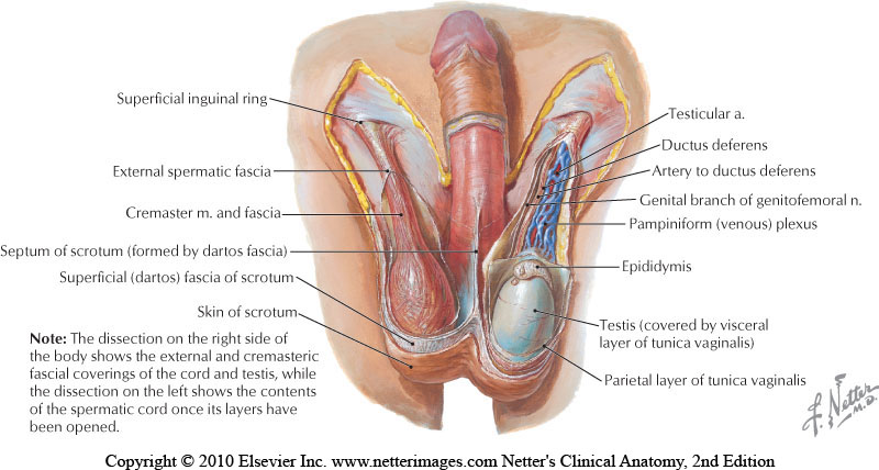
The goal of a vasectomy is typically contraception. The tube known as either the vas deferens or the ductus deferens is interrupted so that sperm does not travel to its typical destination outside the body via the urethra. This disruption in the tube takes place within the spermatic cord as it passes through the scrotum as this area is easily accessible. There are several techniques that can disrupt the tube where the sperm travels including, but not limited to, clamping, cauterization, or excision. The procedure leaves a small incision in the scrotal tissue.
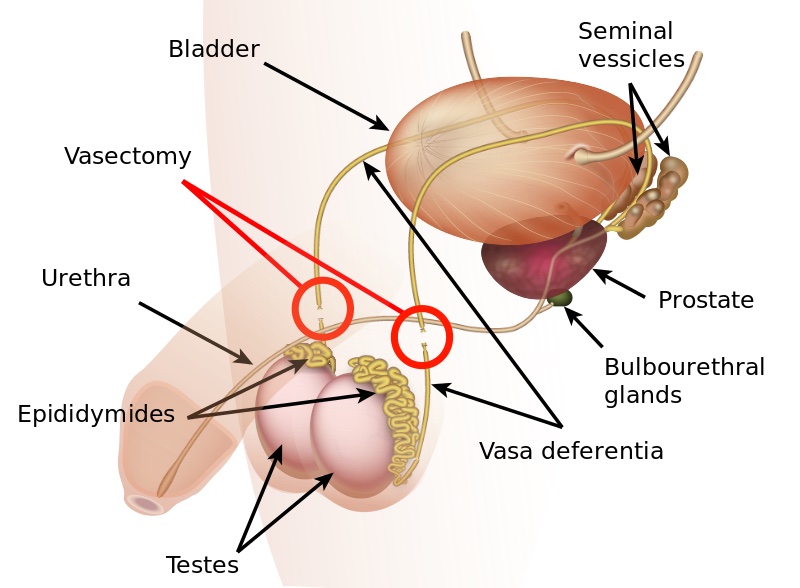
Post-vasectomy Complications
Complications of a vasectomy may include bleeding and hematoma, infection, sperm granuloma (discussed below), chronic scrotal pain, seminal vesicle abscess (rare), and early or late canalization. (Sihra et al., 2007) Interestingly, some patients report less pain after vasectomy. (Leslie et al., 2007) Theories about the cause of post-vasectomy pain include interstitial fibrosis in the epididymal duct and perineurial fibrosis. (Lee et al., 2012) When we consider the anatomy, within the canal there may also be nerve irritation from the genitofemoral nerve, for example, or other connective tissues. If a patient had pain prior to the procedure in the low back, lower abdomen, or groin, the patient’s system may have been vulnerable to complications due to a sensitized system.
Examination & Rehab Efforts
When a patient presents with pain post-vasectomy, symptoms may worsen with prolonged sitting, with pressure from clothing, or in association with sexual or fitness activities. Because there has been a local insult to the tissues, it is logical to check the site of the procedure for any breakdown, signs of significant inflammation, swelling, and to examine for signs of infection such as fever. (Most patients have returned to their medical provider once pain develops, but if they haven’t, a referral is appropriate.) If the pain can be reproduced locally at the site of the procedure, the pain can often be managed by local treatment. You might find benefit in exam procedures such as a trunk or hip extension for the soft tissue tensioning as well as mechanical loading; palpation to the abdominal wall as well as within the spermatic cord. Treatment can address guarding of the area, general wellness (nutrition, movement, mental health), simple modalities such as heat, and gentle self-mobilization to the painful area.
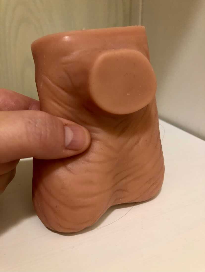
Granulomas
Granulomas can form following a vasectomy, and while usually asymptomatic, a granuloma may be responsible for post-vasectomy pain. They are described as a “bag-like” structure with disintegrating spermatozoa that form at the cut ends of a vasectomy. (Chatterjee et al., 2001) If the granuloma is painful, very light manual mobilization of the thickened area may be done to alleviate pain (see image below). Mobilization of the spermatic cord itself via the testicle or more proximally may also prove helpful. Local modalities such as ultrasound or heat may improve symptoms as well, but clinically I have found that gentle manual therapy and movement exercises are enough to resolve the pain within a few weeks. Patients can be instructed to complete self-mobilization to the area of the granuloma, and as they often are scared to touch the area, helping alleviate this fear is useful in healing.
Post-vasectomy syndrome is very challenging for patients to manage, as they are often dismissed once the procedure is completed. Patients will share that they have been told “everything looks healed” and that the pain should go away on its own. Most providers are unaware of the role of pelvic rehab clinicians, and many pelvic rehab providers are less knowledgeable about conditions related to the scrotum and spermatic cord. For patients who do not respond to conservative intervention, vasectomy reversals have been found to be significantly helpful in reducing pain, though it’s often undesired due to the goal of contraception that inspired the vasectomy. (Herrel et al., 2015; Polackwith et al., 2015). Ideally, patients will be provided with an early recommendation to pelvic rehab so that further procedures or undoing of the vasectomy is avoided.
If you’d like to learn more about post-vasectomy syndrome and many other conditions that can go unrecognized and under-treated, the next opportunity to take the Male Pelvic Floor course is coming up July 9-10,2021!
Chatterjee, S., Rahman, M. M., Laloraya, M., & Pradeep Kumar, G. (2001). Sperm disposal system in spermatic granuloma: a link with superoxide radicals. International journal of andrology, 24(5), 278-283.
Herrel, L. A., Goodman, M., Goldstein, M., & Hsiao, W. (2015). Outcomes of microsurgical vasovasostomy for vasectomy reversal: a meta-analysis and systematic review. Urology, 85(4), 819-825.
Lee, J. Y., Chang, J. S., Lee, S. H., Ham, W. S., Cho, H. J., Yoo, T. K., ... & Lee, S. W. (2012). Efficacy of vasectomy reversal according to patency for the surgical treatment of postvasectomy pain syndrome. International journal of impotence research, 24(5), 202-205.
Leslie, T.A., R.O. Illing, D.W. Cranston, J. Guillebaud (2007). “The incidence of chronic scrotal pain after vasectomy: a prospective audit.” BJU International 100: 1330-1333.
Polackwich, A. S., Tadros, N. N., Ostrowski, K. A., Kent, J., Conlin, M. J., Hedges, J. C., & Fuchs, E. F. (2015). Vasectomy reversal for postvasectomy pain syndrome: a study and literature review. Urology, 86(2), 269-272.

Herman & Wallace currently has satellite courses, remote courses, and online courses offered through our partner, Medbridge. These online courses provide education and patient engagement tools for pelvic floor dysfunction. H&W faculty have put together a collection of online continuing education courses with our partners at MedBridge. These convenient learning resources and can be purchased individually or as part of an annual subscription
The truth is that we all have hectic busy schedules that can make setting aside time for a live course can be difficult. Annual subscribers get access to all 800+ courses on the Medbridge site, their Home Exercise Program, and Patient Engagement platforms! As a Herman & Wallace referral, you are eligible for a discounted subscription with access to all of the content at MedBridge with promo code HWoverview.
Heather Rader, PT, DPT, PRPC, BCB-PMD recorded a new series for Medbridge last summer. Her courses explore:
- anatomical relevance of the pelvic girdle and pelvic floor structures
- common movement dysfunctions in rehabilitation
- overview of the sacroiliac joint, pubic symphysis, and coccyx
- bladder, bowel, and sexual dysfunctions created by pelvic floor dysfunctions
- connections between orthopedic dysfunction seen with pelvic floor dysfunctions
- orthopedic conditions such as back pain, hip pain, orthopedic trauma, and surgery
- diastasis recti abdominis cluster with pelvic floor dysfunctions, such as incontinence and pelvic pain syndromes in clinical practice
- including several other topics
The Medbridge online course catalog contains an in-depth physical therapy section with many familiar instructors including Holly Tanner and Steven Dischiavi. These online courses are a great way for pelvic rehabilitation specialists to gain knowledge and skills and complete CEU requirements without having to commit to a predetermined schedule.
Herman & Wallace continuing education courses are developed and instructed by our nationally recognized faculty members. Contact hours vary by course and may apply towards continuing education requirements according to each state.

The following is our interview with Jazma Dobbins PT, DPT, PRPC, CAPP-Pelvic. Jazma recently passed the Pelvic Rehabilitation Practitioner Certification (PRPC) exam. She practices in at TherapySouth in Gadsen, AL and is a Teaching Assistant for local Alabama satellite courses with H&W. Jazma was kind enough to share some thoughts about her career with us. Thank you, Jazma - and congratulations on receiving your PRPC!
Q: How did you get involved in the pelvic rehabilitation field?
A: Educating others about ways to empower themselves through healthcare advocacy is extremely important to me. I knew I wanted to be a pelvic health physical therapist the moment I read a similarly titled article in a women’s health magazine over 10 years ago. I was experiencing painful intercourse and urge incontinence. I was 20 years old and felt ashamed, abnormal, and alone. I had been given the unfortunate advice of “try drinking wine to relax” and “never drink alcohol if you have urge incontinence." So many inconsistencies to my young and frustrated mind.
That is when it dawned on me that I could do that, I could be a pelvic health physical therapist. Then we would have that service in my community. I was certain, and rightly so, that there were plenty of other women (I did not yet know of men’s health issues) who needed these services.
Q: What has your educational journey as a pelvic rehab therapist looked like?
A: I started my pelvic rehab education journey as a third-year student in PT school. I took the pelvic health level 1 course through the then called Section on Women’s Health. Needless to say, I was hooked on pelvic rehab. Over the following year, I completed the APTA pelvic series and earned my CAPP-Pelvic. In 2019 I did the Coccydynia and painful sitting course through Herman and Wallace and fell in love with H&W! Since then, I have had the pleasure of being a Teaching Assistant for multiple H&W courses.
Q: What do you love about assisting at courses?
A: What I love most about serving as a teaching assistant is the opportunity to learn from the participants. I have yet to TA a course where I didn’t learn a new skill or thought process. I love to TA the pelvic level 1 course most of all because of the eagerness and anticipation around learning a whole new world of what PT can do for our clients. I love to spread energy and passion and help create an environment of truly nonjudgmental learning.
Q: What is your message to course participants who are just starting their journey?
A: Keep pursuing your passion! Let no learning opportunity go unutilized and never stop learning!

This blog contains excerpts from an interview with Tara Sullivan, PT, DPT, PRPC, WCS, IF. Tara started in the healthcare field as a massage therapist, practicing over ten years including three years of teaching massage and anatomy and physiology. Tara has specialized exclusively in Pelvic Floor Dysfunction treating bowel, bladder, sexual dysfunctions, and pelvic pain since 2012.
Acute pain can indicate specific injury to the body. Chronic pain is very different. With Chronic Pelvic Pain (CPP) the initial injury has healed, but the pain continues because of changes in the nervous system, muscles, and tissues. Recognizing that the nervous system influences pain perception, especially in the chronic pelvic pain population, is the first step in treating these patients, but is it enough? Tara Sullivan and Alyson Lowrey are presenting a new remote course on chronic pelvic pain called Pain Science for the Chronic Pelvic Pain Population scheduled for July 17-18, 2021.
The medical definition of pain is an unpleasant feeling that is conveyed to the brain by sensory neurons. Pain is a universal experience that serves to alert the brain to potential damage to the body. It performs the function of triggering avoidance to preserve itself from harm. Oddly, the strength and unpleasantness of pain is not directly related to the nature or extent of the damage.
When the pain signal remains active in the nervous system for longer than six months and persists after the triggering event has healed, then it is cataloged as chronic pain. There is another layer when experiencing chronic pain known as central sensitization. This is an increased responsiveness of the nervous system that results in hypersensitivity and an increased pain response outside the area of injury. Pain itself can produce systematic and chemical brain changes resulting in more pain from fewer stimuli.
The course, Pain Science for the Chronic Pelvic Pain Population, offers tools to recognize when sensitization may be playing a role and provides the framework needed to apply pain science to the chronic pelvic pain population. In this course, you will gain an understanding and expand your knowledge on how pain science specifically presents in patients suffering from endometriosis, interstitial cystitis, primary dysmenorrhea, pelvic floor muscle overactivity, vulvodynia/vestibulodynia, vaginismus, and prostatitis.
Case studies and specific intervention techniques, including how to explain pain to a patient, are discussed so participants leave with the confidence to address the missing link in treating your patient’s chronic pelvic pain. We will also discuss how common rehab interventions such as manual therapy, dry needling, biofeedback, graded exposure, and therapeutic exercise assist in downregulating the nervous system.
On July 17-18th, 2021, Alyson Lowrey and Tara Sullivan team up to give you their combined experience of orthopedic and pelvic health in treating this population in the course Pain Science for the Chronic Pelvic Pain Population.

This blog contains excerpts from an interview with Pamela A. Downey, PT, DPT, WCS, BCB-PMD, PRPC, Pamela is a Board Certified Clinical Specialist in Women’s Health Physical Therapy and Board Certified in Biofeedback for Pelvic Muscle Dysfunction. She is the owner of Partnership in Therapy, private practice in Coral Gables, Florida. Dr. Downey's treatment focuses are pelvic floor dysfunction, urogynecological and colorectal issues, spine dysfunction, osteoporosis, and complaints associated with pregnancy and postpartum. Her mission is to educate and integrate healthy lifestyles for patients on the road to wellness.
Physical therapists often require special training to treat pudendal neuralgia. Pamela A. Downey is partnering with H&W to teach the Pudendal Neuralgia and Nerve Entrapment Remote Course, scheduled for June 19-20, 2021. This course teaches pudendal neuralgia diagnostic skills for practitioners to have an improved impact in treating patients with pudendal nerve/pelvic floor muscle dysfunctions.
Pudendal neuralgia is also known as Alcock’s syndrome, pudendal canal syndrome, or cyclist syndrome. This condition is caused by tension, compression, or entrapment of the pudendal nerve, and leads to pelvic pain, sexual dysfunction, difficulty with urination and defecation, among other issues.
Pudendal neuralgia is often unrecognized by physicians, including gynecologists, urologists, and neurologists. Dr. Downey observes that “Organizing your clinical decision-making process is key in determining the source of seated pain. Pudendal neuralgia can be a chicken and egg clinical phenomenon. My success comes from relying on a solid anatomy background in helping solve the pudendal puzzle.”
Successful treatments can include connective tissue mobilization, neural mobilization, and a home exercise program. Poor movement patterns can contribute to the symptoms of pudendal neuralgia. Physical therapy evaluation in these cases can include movement assessment and a gentle internal assessment of the patient's pelvic muscles. This provides information about the muscles’ ability to contract and relax. Exercises recommended to relax the pudendal nerve and provide temporary relief include cobra pose, side-lying hip abduction and extension, and wide-leg bridges.
Dr. Downey shares that she loves teaching the Pudendal Neuralgia and Nerve Entrapment Remote Course. “We teach the participants, in real-time, how to use evidence-based criteria to see if pudendal neuralgia makes sense as the driving diagnosis. Then we develop this confidence by careful dissection of case studies of real patients treated out over multiple visits, just like you do in the clinic."
Hone your decision-making process and gain confidence in the Pudendal Neuralgia and Nerve Entrapment Remote Course to treat pelvic pain with Pamela Downey on June 19-20, 2021.

Working with Physiatry for Pelvic Pain is a new remote course created by Dr. Allyson Shrikhande, scheduled for Jun 27, 2021. This course overviews the synergistic nature of pelvic physiatry with pelvic floor physical therapy, in hopes of promoting collaboration for the care of male and female chronic pelvic pain patients.
Dr. Allyson Shrikhande is a board-certified Physical Medicine and Rehabilitation specialist and is the Chair of the Medical Education Committee for the International Pelvic Pain Society. Allyson has published peer-reviewed articles on the treatment of muscle pain in academic journals and works closely with renowned pelvic pain gynecologists and urologists. Taking a team approach, she works with specialists in pelvic floor physical therapy, kinetics and movement, as well as acupuncturists, nutritionists, cognitive-behavioral therapists, and functional medicine physicians.
The following is our interview with Allyson Shrikhande on physiatry.
Q: What is a physiatrist?
A: A physiatrist is an MD or DO with a specialty in Physical Medicine and Rehabilitation. This non-operative medical discipline involves focusing on the neuromusculoskeletal system to help patients recover their functional well-being and quality of life. We describe physiatry as an extension of physical therapy because a physiatrist diagnoses, manages, and treats pain from injury, illness, or medical conditions, incorporating other methods in concert with physical therapy to rehabilitate the body. Physiatrists are trained not solely in one organ system – rather, they take a holistic, full-body approach that accounts for the interplay of different organ systems, both with each other and with the neuromuscular and myofascial systems.
Q: What does a physiatrist do?
A: Physiatrists work with physical therapy to rehabilitate the neuromuscular system. A core underlying theme in physiatry is the concept of Neuroplasticity. This is the understanding that the nervous system has the ability to form and reorganize synaptic connections, especially in response to experience or learning following injury.
Q: What do physiatrists treat?
A: Because physiatrists focus on the interconnected systems of the entire body, they treat a wide range of injuries and disorders. Physiatrists commonly work with patients who have pelvic, back or neck pain who are recovering from issues such as sports injuries, neuromuscular disorders, arthritis, or injuries to the brain or spinal cord.
Q: Why would I see a physiatrist?
A: At Pelvic Rehabilitation Medicine, our Pelvic Physiatrists diagnose and treat the structures of the pelvis – the muscles, nerves, and joints. One of our physiatrists can provide non-operative options to medically manage and treat pelvic pain and pelvic floor muscle dysfunction. We treat an array of symptoms under the umbrella of pelvic pain which includes pain with intercourse, urinary urgency/frequency or pain with urination, constipation or painful bowel movements, and pain affecting the coccyx, groin, pelvis, lower back, and lower abdomen.
Q: As a pelvic floor physical therapist, what can I learn from a physiatrist?
A: The relationship between physiatry and physical therapy is vital to the collaborative approach that our pelvic pain patients require. Physiatrists perform a full neuromuscular exam (including an internal pelvic floor exam) and can order imaging, prescribe oral medications, suppositories, and topical medications for some patients. Our physiatrists can also perform safe outpatient ultrasound-guided procedures to treat underlying neuromuscular dysfunction, all in combination with continued pelvic floor PT when appropriate.
By accepting you will be accessing a service provided by a third-party external to https://hermanwallace.com/
All Upcoming Continuing Education Courses


Mobilization of Gastrointestinal Visceral Fascia Satellite Lab Course - Self-Hosted - December 6 - 8 2024
Dec 6 2024 - Dec 8 2024

Mobilization of Visceral Fascia: The Gastrointestinal System - Phoenix AZ - December 6 - 8 2024
Dec 6 2024 - Dec 8 2024

Mobilization of Visceral Fascia: The Gastrointestinal System Satellite Lab Course - Woodbury NY - December 6 - 8 2024 - SOLD OUT
Dec 6 2024 - Dec 8 2024

Mobilization of Visceral Fascia - Gastrointestinal Satellite Lab Course - Raleigh NC - December 6 - 8 2024
Dec 6 2024 - Dec 8 2024

Mobilization of Visceral Fascia - Gastrointestinal Satellite Lab Course - Medford OR - December 6 - 8 2024
Dec 6 2024 - Dec 8 2024

Mobilization of Visceral Fascia: Gastrointestinal Satellite Lab Course - Milwaukee WI - December 6 - 8 2024
Dec 6 2024 - Dec 8 2024

Mobilization of Visceral Fascia - Gastrointestinal Satellite Lab Course - Ann Arbor MI - December 6 - 8 2024
Dec 6 2024 - Dec 8 2024

Nutrition Perspectives for the Pelvic Rehab Therapist - Remote Course - December 7 - 8 2024
Dec 7 2024 - Dec 8 2024
Biofeedback for Pelvic Muscle Dysfunction Satellite Lab Course - Self-Hosted - December 7 2024
Dec 7 2024

Sacral Nerve Manual Assessment and Treatment - Remote Course - December 7 - 8 2024
Dec 7 2024 - Dec 8 2024


Pelvic Function Level 2B - Satellite - Colorado Springs CO - December 7 - 8 2024
Dec 7 2024 - Dec 8 2024


Pelvic Function Level 1 - Satellite - Denver CO - December 14 - 15 2024 - SOLD OUT
Dec 14 2024 - Dec 15 2024
Pelvic Function Level 1 - Satellite Lab Course - Lone Tree CO - December 14 - 15 2024 - SOLD OUT
Dec 14 2024 - Dec 15 2024

Pelvic Function Level 1 - Satellite - Jacksonville FL - December 14 - 15 2024
Dec 14 2024 - Dec 15 2024

Pelvic Function Level 1 - In-Person - New York NY - December 22 - 23 2024 - SOLD OUT
Dec 22 2024 - Dec 23 2024


Pelvic Function Level 1 - In-Person - Philadelphia PA - January 11 - 12 2025 - SOLD OUT
Jan 11 2025 - Jan 12 2025

Oncology of the Pelvic Floor Level 1 - Remote Course - January 11 - 12 2025
Jan 11 2025 - Jan 12 2025

Modalities and Pelvic Function - In-Person - Sacramento CA - January 11 - 12 2025
Jan 11 2025 - Jan 12 2025

Pediatrics Level 1 - Treatment of Bowel and Bladder Disorders - Remote Course - January 11 - 12 2025
Jan 11 2025 - Jan 12 2025


Pelvic Function Level 1 - Satellite - Knoxville TN - January 18 - 19 2025 - SOLD OUT
Jan 18 2025 - Jan 19 2025


Pelvic Function Level 1 - Satellite - Manchester IA - January 18 - 19 2025
Jan 18 2025 - Jan 19 2025

Pelvic Function Level 1 - Satellite - Long Beach CA - January 18 - 19 2025
Jan 18 2025 - Jan 19 2025


Pelvic Function Level 1 - Satellite - Fort Lauderdale FL - January 18 - 19 2025
Jan 18 2025 - Jan 19 2025

Pelvic Function Level 2B - Satellite - Bradenton FL - January 18 - 19 2025
Jan 18 2025 - Jan 19 2025
Pelvic Function Level 2B - Satellite - Commerce Township MI - January 18 - 19 2025
Jan 18 2025 - Jan 19 2025