Upcoming Courses Near You
Finding courses near you...

Pelvic Rehab Report
The official Herman & Wallace blog. New blogs post every Friday on topics relating to the field of pelvic floor dysfunction.

Male pelvic cancer survivors, including those treated for prostate, bladder, penile, and testicular cancers, face a range of ongoing functional challenges that affect quality of life. Treatments such as radical prostatectomy, radiation therapy, chemotherapy, and reconstructive surgery can lead to urinary incontinence, erectile dysfunction, bowel irregularity, pelvic pain, and changes in body image or hormonal balance. These effects often overlap and require an integrated rehabilitation approach.
Prostate cancer survivors frequently report urinary leakage, urgency, and reduced bladder control. Bladder cancer patients, particularly those who undergo urinary diversion or neobladder reconstruction, may struggle with altered storage and emptying patterns. Colorectal cancer survivors treated with low anterior resection often develop low anterior resection syndrome, which is characterized by stool clustering, urgency, and incontinence (Jones et al., 2024; Kim & Oh, 2023). Penile and testicular cancer survivors may experience sexual dysfunction and hormonal disruptions that influence pelvic floor function and psychosocial well-being. Across these diagnoses, common contributors to dysfunction include pelvic floor muscle weakness or discoordination, neural disruption, scar tissue and fibrosis, and the cumulative impact of cancer treatment on mobility, confidence, and daily function.
The Role of Pelvic Floor Rehabilitation
Pelvic floor rehabilitation is an essential component of survivorship care. Randomized trials and systematic reviews consistently support structured pelvic floor muscle training to improve urinary continence after prostatectomy (Fernández et al., 2015; Chen et al., 2023; Gerlegiz et al., 2025). Supervised PFMT produces better outcomes than unsupervised exercise, particularly when initiated before surgery or early postoperatively. Confirming accurate pelvic floor activation through biofeedback or palpation is critical for optimizing treatment success (Gerlegiz et al., 2025).
For men treated with pelvic radiation, long-term changes in muscle structure and neural control can contribute to urinary or bowel dysfunction and pelvic pain. Morphological and functional assessments using MRI, surface electromyography, and palpation have demonstrated reduced pelvic floor muscle endurance and altered activation patterns years after treatment (Ribeiro et al., 2021). These findings highlight the importance of ongoing rehabilitation to restore motor control, manage fibrosis, and reduce symptom burden.
Colorectal cancer survivors with low anterior resection syndrome benefit from targeted pelvic floor rehabilitation. Structured programs including pelvic floor muscle training, coordination exercises, and biofeedback have demonstrated improvements in bowel function, urgency, and quality of life (Jones et al., 2024; Kim & Oh, 2023). Programs delivered over multiple sessions, with patient adherence to home exercises, provide the most consistent benefit.
Sexual health is another domain where pelvic rehabilitation is important. Pelvic floor muscle training can improve erectile function, particularly when initiated preoperatively or in high-volume programs (Milios et al., 2020; Wong et al., 2020). Pelvic therapists can also address pelvic pain, scar sensitivity, and coordination deficits that contribute to sexual discomfort. Multidisciplinary collaboration with urology, sexual medicine, and mental health professionals provides comprehensive support for survivors navigating intimacy and relationship challenges.
Practical Rehabilitation Strategies Evidence-based rehabilitation for male pelvic cancer survivors should include the following components:
Assessment and activation - Confirm voluntary pelvic floor contraction using digital palpation, biofeedback, or ultrasound. Accurate assessment allows for individualized exercise prescription and objective tracking of progress (Gerlegiz et al., 2025; Ribeiro et al., 2021).
Structured exercise prescription - High-volume pelvic floor muscle training incorporating both slow- and fast-twitch fibers is recommended. Supervised sessions ensure correct technique, increase adherence, and improve outcomes (Fernández et al., 2015; Chen et al., 2023).
Biofeedback and adjunct modalities - Biofeedback supports motor learning and awareness. Electrical stimulation may be used selectively in patients unable to contract muscles effectively (Fernández et al., 2015).
Bowel retraining and coordination exercises - Strategies such as urge suppression, stool consistency management, scheduled toileting, and coordination exercises improve function in patients with low anterior resection syndrome (Jones et al., 2024; Kim & Oh, 2023).
Manual therapy and scar management - Hands-on techniques address fibrosis, scarring, restricted mobility, and pain. Scar desensitization and soft tissue mobilization support improved muscle recruitment and pelvic comfort.
Sexual rehabilitation integration - Pelvic floor muscle training can complement medical penile rehabilitation, graded exposure, and sensory retraining to improve sexual function and comfort (Milios et al., 2020; Wong et al., 2020).
Psychosocial support - Addressing body image, anxiety, and intimacy concerns is essential. Counseling referrals and supportive communication improve adherence and quality of life.
Prehabilitation and telehealth - Preoperative pelvic floor training can improve postoperative outcomes (Chen et al., 2023). Telehealth facilitates remote guidance, adherence monitoring, and access to specialized pelvic rehabilitation services.
Takeaway
Functional impairments after male pelvic cancer treatment are common, but rehabilitation can significantly improve urinary continence, bowel control, sexual function, pain, and overall quality of life. Evidence strongly supports structured, supervised pelvic floor muscle training for urinary incontinence after prostatectomy, with growing support for bowel and sexual rehabilitation in this population. Early, individualized, and evidence-based intervention is key to maximizing recovery.
Clinicians seeking to deepen their skills in treating male pelvic and colorectal cancer survivors are encouraged to register for the Oncology of the Pelvic Floor Level 2A: Male Pelvic and Colorectal Cancers course on December 6–7. This two-day training provides hands-on learning, case-based discussions, and practical strategies for evidence-based assessment and intervention.
References
- Fernández RA, García-Hermoso A, Solera-Martínez M, Correa MT, Morales AF, Martínez-Vizcaíno V. Improvement of continence rate with pelvic floor muscle training post-prostatectomy: a meta-analysis of randomized controlled trials. Urol Int. 2015;94(2):125-32. doi: 10.1159/000368618. PMID: 25427689.
- Ribeiro AM, Nammur LG, Mateus-Vasconcelos ECL, Ferreira CHJ, Muglia VF, de Oliveira HF. Pelvic floor muscles after prostate radiation therapy: morpho-functional assessment by magnetic resonance imaging, surface electromyography and digital anal palpation. Int Braz J Urol. 2021 Jan-Feb;47(1):120-130. doi: 10.1590/S1677-5538.IBJU.2019.0765. PMID: 33047917; PMCID: PMC7712707.
- Milios JE, Ackland TR, Green DJ. Pelvic Floor Muscle Training and Erectile Dysfunction in Radical Prostatectomy: A Randomized Controlled Trial Investigating a Non-Invasive Addition to Penile Rehabilitation. Sex Med. 2020 Sep;8(3):414-421. doi: 10.1016/j.esxm.2020.03.005. Epub 2020 May 14. PMID: 32418881; PMCID: PMC7471070.
- Chen, Yi-Hsuan1; Juan, Yung-Shun1,2,3,4; Wei, Wei-Chi1; Geng, Jiun-Hung5; Chueh, Kuang-Shun1,4; Lee, Hsiang-Ying1,2,3,4,*. Effects of Early Pelvic Floor Muscle Training on Early Recovery of Urinary Incontinence after Prostate Surgery. Urological Science 34(1):p 39-45, Jan–Mar 2023. | DOI: 10.4103/UROS.UROS_59_22
- Wong C, Louie DR, Beach C. A Systematic Review of Pelvic Floor Muscle Training for Erectile Dysfunction After Prostatectomy and Recommendations to Guide Further Research. J Sex Med. 2020 Apr;17(4):737-748. doi: 10.1016/j.jsxm.2020.01.008. Epub 2020 Feb 3. PMID: 32029399.
- Jones S, Edie A, Troop E, Hill JS, Thompson JA. The Effect of Pelvic Floor Rehabilitation on Low Anterior Resection Syndrome After Colorectal Cancer Treatment. J Adv Pract Oncol. 2024 May 22:1-12. doi: 10.6004/jadpro.2024.15.8.4. Epub ahead of print. PMID: 39802528; PMCID: PMC11715399.
- Kim YM, Oh EG. Effectiveness of Pelvic Floor Muscle Training for Patients Following Low Anterior Resection: A Systematic Review and Meta-analysis. J Wound Ostomy Continence Nurs. 2023 Mar-Apr 01;50(2):142-150. doi: 10.1097/WON.0000000000000958. PMID: 36867038.
- Gerlegiz ENA, Öztürk D, Gürşen C, Akbayrak T, Özgül S. Structured and supervised pelvic floor muscle training following confirmed contraction in post-prostatectomy urinary incontinence: a systematic review of randomized controlled trials. J Cancer Surviv. 2025 Aug 28. doi: 10.1007/s11764-025-01882-6. Epub ahead of print. PMID: 40877548.

Pelvic rehabilitation practitioners often focus on mechanical concerns such as diastasis recti, pelvic floor weakness, and altered load transfer. However, one foundational element that deserves equal attention in the postpartum period is the ongoing effect of hormonal shifts on connective tissues and joints.
Even after delivery, hormones such as relaxin, estrogen, and progesterone continue to influence tissue behavior, joint mobility, and the body's response to rehabilitation. Understanding how these hormones affect ligament laxity, collagen turnover, and neuromuscular control helps clinicians design safer and more effective recovery programs.
Hormonal Changes in Pregnancy and Early Postpartum
Relaxin is produced by the corpus luteum and placenta, and rises significantly during pregnancy. It is known for its role in “relaxing” muscles and ligaments, especially in the pelvis, to facilitate childbirth. Estrogen and progesterone also rise and modulate connective-tissue metabolism and receptor expression in ligaments.
A recent review highlights that pregnancy-associated hormonal fluctuations (relaxin, estrogen, progesterone) contribute to increased joint laxity (Yalçınkaya et al., 2025). These hormonal effects do not cease abruptly at delivery; they taper variably and may persist for months postpartum, meaning joint and tissue behavior remains altered during rehab.
Hormones Affect Connective Tissue and Joint Mechanics
At the cellular level, relaxin increases the activity of matrix metalloproteinases (MMPs) such as MMP-1, -9, and -13, which degrade collagen and weaken ligament/tendon architecture (Parker et al., 2-22). Estrogen and progesterone increase expression of relaxin receptors in ligaments, amplifying these effects (Yalçınkaya et al., 2025).
In animal and human tendon studies, both relaxin and estrogen have been shown to reduce tendon stiffness and increase compliance, which can reduce load tolerance (Danos et al., 2023). The net mechanical effect is increased joint mobility (or perceived laxity), reduced passive stability, and a greater need for neuromuscular control to compensate.
Clinically, this manifests as more “loose-feeling” joints, increased cushioning of movement, greater reliance on muscular control for stability, and potentially slower progress of load-transfer training. A prospective cohort during pregnancy found associations between estrogen changes and increased low-back/pelvic-girdle pain and disability (Daneau et al., 2025). Although postpartum longitudinal data are sparse, the same mechanisms are likely to persist into the early postpartum period and influence rehab.
Implications for postpartum rehabilitation of joints
- Assessment and monitoring: Include joint laxity screening (e.g., Beighton score or joint-specific tests) and ask the client about subjective joint “giveness” or instability. Recognize that normal endpoints may be slower to return.
- Loading progression: Because connective tissues are in a transiently weakened state, adopt more conservative load progressions. Emphasize neuromuscular control (motor control, stability) before heavy mechanical loading. Use subsets such as closed-chain, low-velocity, high-control activities.
- Tailored timeline: Do not assume pre-pregnancy tissue behavior has returned simply because the organ-based recovery period has ended. A busy schedule, hormonal fluctuations (including lactation, menses return) may continue to modulate tissue mechanics.
- Patient education: Explain that “it’s not just about the tummy and pelvic floor” but that “your ligaments and joints are still adjusting.” This can help set realistic expectations and foster adherence to graded rehab.
- Coordination with other practitioners: Ensure communication with obstetric, primary-care, and sports-medicine colleagues about longer-term musculoskeletal implications of hormonal and biomechanical changes. As one review concluded, many women are at elevated risk for persistent joint instability and possible long-term sequelae such as early osteoarthritis (Yalçınkaya et al.., 2025).
Case example
A 34-year-old primipara at 10 weeks postpartum presents with “clicking” in the pubic symphysis region when lifting her 9-month-old toddler and reports a sense of “unstable hips” when stepping sideways. On assessment, she has a Beighton score of 5/9 (with bilateral thumb-to-forearm and elbow hyperextension). She also reports previous hip discomfort in adolescence.
Given her history and findings, you design a graduated program: phase 1 focused on pelvic-floor activation + hip stability in non-weight-bearing by week 12; phase 2 at week 16, introducing unilateral step-downs with low amplitude; phase 3 at week 20, adding higher load functional tasks (carrying child + step).
You monitor joint symptoms, ensure neuromuscular control precedes full load, and educate her regarding ligamentous recovery timeline (~6-12 months). You explain that although the baby is 9 months old, her connective tissues may still be adjusting to hormone-mediated changes.
Conclusion
Hormonal recovery is a critical but sometimes overlooked element of postpartum rehabilitation. The lingering influence of relaxin, estrogen, and progesterone shapes how connective tissues behave and respond to loading. By integrating hormonal awareness into clinical decision-making, pelvic health practitioners can enhance precision, promote safety, and improve long-term functional outcomes. Recovery after childbirth is not limited to muscle or fascia; it is a systemic process involving hormones, tissues, and time.
For clinicians interested in expanding their postpartum rehabilitation skills, consider registering for the upcoming Postpartum Rehabilitation Remote Course scheduled for December 13-14. This course covers acute postpartum management, mental health screening, and musculoskeletal considerations. Participants will learn to modify examinations and interventions for the relevant stages of postpartum recovery. In addition to abdominal wall considerations, typical spine and extremity dysfunctions will be addressed. The course includes instruction on postpartum exercise and return to fitness, with labs covering external perineal screening as well as techniques for the abdominal wall, spine, and ribs, and upper and lower quarter dysfunction.
References
- Yalçınkaya, B., Sezgin, E. A., Saçıntı, K. G., & Özçakar, L. (2025). Neuromusculoskeletal disorders in pregnancy revisited. Journal of Joint Diseases and Related Surgery, 36(3), 741-750.
- Parker, E. A., Meyer, A. M., Goetz, J. E., Willey, M. C., & Westermann, R. W. (2022). Do Relaxin Levels Impact Hip Injury Incidence in Women? A Scoping Review. Frontiers in Endocrinology, 13, 827512. https://doi.org/10.3389/fendo.2022.827512
- Danos, N., Patrick, M., Barretto, J., Bilotta, F., & Lee, M. (2023). Effects of pregnancy and lactation on muscle-tendon morphology. Journal of Anatomy, 243(5), 860-869. https://doi.org/10.1111/joa.13916
- Daneau, C., Nougarou, F., Abboud, J., Ruchat, M., & Descarreaux, M. (2025). Changes in pregnancy-related hormones, neuromechanical adaptations and clinical pain status throughout pregnancy: A prospective cohort study. PLOS ONE, 20(2), e0314158. https://doi.org/10.1371/journal.pone.0314158

In a recent article entitled “Physiotherapy Approaches in Individuals with Functional Defecation Disorders: Literature Review,” published in the Journal of Health Sciences in August of 2025, the use of surface EMG biofeedback (sEMG) was recommended as a first-step treatment for functional defecation disorders. It was concluded that biofeedback applications were superior to standard treatment and effective in improving patients’ quality of life. Both the American Neurogastroenterology and Motility Society and the American College of Gastroenterology recommend biofeedback as first-line therapy for dyssynergic defecation.
Surface electromyography (sEMG) biofeedback has emerged as one of the most effective conservative interventions for treating dyssynergic defecation, also known as pelvic floor dyssynergia. This condition is characterized by the paradoxical contraction or inadequate relaxation of the pelvic floor muscles and external anal sphincter during attempted defecation, resulting in functional outlet obstruction and chronic constipation. Dyssynergia affects many of our clients with chronic constipation and is a prevalent referral diagnosis in pelvic floor rehabilitation. For pelvic health therapists, sEMG biofeedback provides a valuable therapeutic tool that directly retrains neuromuscular coordination, corrects maladaptive defecation mechanics, and improves patient outcomes without the risks associated with pharmacologic or surgical interventions.
How Surface EMG Biofeedback Works
Biofeedback therapy addresses the underlying pathophysiology of dyssynergia by helping patients learn to modulate pelvic floor muscle activity during simulated defecation. Surface EMG biofeedback uses external perianal or intrarectal sensors to record muscle activation patterns, visually displaying motor unit firing in real time on a monitor. When patients attempt defecation, those with dyssynergia often engage in compensatory or dysfunctional mechanics such as breath-holding, strain maneuver without pelvic floor relaxation, or tightening of the external anal sphincter and puborectalis muscle. Biofeedback enables patients to see these abnormalities and receive immediate coaching to correct them. This learning model aligns with the principles of motor relearning and neuroplasticity and is particularly effective for reeducating muscles involved in autonomic and habitual reflex responses, including those of the pelvic floor.
A key benefit of surface EMG biofeedback is its ability to correct maladaptive motor patterns rather than merely provide symptomatic relief. Unlike laxatives, which simply soften stool or increase motility, biofeedback teaches coordinated defecation by training patients to relax the anal sphincter, lengthen the pelvic floor, and generate appropriate intra-abdominal pressure. Many patients demonstrate dyssynergic patterns such as Type I dyssynergia, where the pelvic floor contracts during attempted evacuation, or Type III, where inadequate abdominal force is generated. Biofeedback helps individualize treatment, as therapists can use EMG data to identify specific coordination deficits and modify training accordingly. In addition, EMG signals provide objective progress markers, reinforcing patient engagement and compliance.
Another important clinical advantage of biofeedback is that it is non-invasive, safe, and well-tolerated across patient populations. It is appropriate for adults, older adults, and even motivated pediatric patients. Side effects are minimal, especially with surface EMG sensors that eliminate the need for internal probes when preferred clinically. This makes EMG biofeedback a valuable option for patients who may not tolerate rectal balloon training or invasive anorectal procedures due to pain, anxiety, or trauma history.
Integrating Biofeedback into Pelvic Rehabilitation
Biofeedback therapy integrates well within holistic pelvic rehabilitation programs. Treatment typically includes coordination training, diaphragmatic breathing, colonic massage instruction, toileting posture education, bowel mechanics retraining, and behavioral strategies to restore normal recto-anal sensory reflexes. Surface EMG enhances the effectiveness of behavioral training by improving patients’ proprioceptive awareness of their pelvic floor, a region in which neuromuscular awareness is typically poor. Additionally, EMG biofeedback can incorporate downtraining techniques to reduce high resting pelvic floor tone.
Despite its proven benefits, access to biofeedback therapy can be inconsistent due to limited awareness among healthcare providers and insufficient training opportunities for therapists. However, pelvic health therapists are uniquely positioned to deliver this intervention effectively due to their expertise in neuromuscular reeducation, movement analysis, and behavioral coaching. Incorporating sEMG biofeedback into a pelvic rehabilitation practice not only aligns with evidence-based care but also enhances clinical credibility and expands therapeutic capabilities, especially for patients with chronic functional bowel disorders.
Surface EMG biofeedback offers substantial therapeutic benefits for the treatment of pelvic floor dyssynergia. It is superior to standard medical therapy, targets the root cause of dyssynergia through neuromuscular retraining, promotes long-term bowel function normalization, and empowers patients to take an active role in their recovery. With growing clinical validation and patient demand for non-pharmacologic treatments, sEMG biofeedback should be considered an essential component of pelvic floor therapy practice. For pelvic health therapists seeking to deliver advanced, evidence-based care, biofeedback represents a powerful and effective intervention with lasting results.

Excessive pelvic floor activity during attempts at bowel evacuation creates a mechanical barrier to evacuation and demonstrates dyssynergia (incoordination). When a patient “bears down” the abdominals (blue) should increase, and the pelvic floor (green) should relax.

Correct pattern: Pelvic floor muscles relax and abdominal muscles contract to increase intra-abdominal pressure for proper bowel movement coordination.
Conclusion
A long history of scientific evidence supports the use of sEMG biofeedback for managing incontinence or pain symptoms. As a noninvasive, cost-effective, and powerful treatment modality, healthcare providers should consider this tool when managing pelvic floor dysfunction. Providers should be adequately educated in this valuable modality to make the most of the skills and knowledge gained through this intervention. For more information regarding courses and certification, please visit www.pelvicfloorbiofeedback.com.
Biofeedback for Pelvic Muscle Dysfunction is available in satellite and self-hosted formats.
Biofeedback for Pelvic Muscle Dysfunction, scheduled for December 13, 2025, is led by board-certified Instructors Jane Kaufman and Tiffany Lee, who introduce participants to the use of biofeedback in the treatment of bladder, bowel, and pelvic floor disorders. In this course, participants learn about surface EMG biofeedback by using the equipment on themselves to experience dynamic muscle assessment in supine, sitting, and standing positions. This dynamic course also includes behavioral strategies to relearn proper muscle control and improve pelvic floor function.
If you attend this course at a scheduled hosted location (NOT self-hosted), then the equipment will be supplied by our biofeedback vendor, Current Medical Technologies. Satellite Locations for December 13th are:
Self-hosted participants do not need partners and can treat themselves using the equipment. Equipment used in this course, and required for all self-hosted registrants, is as follows (all items can be purchased on www.cmtmedical.com):- A 2-channel sEMG biofeedback hand-held unit with software on a laptop or computer is required. The instructors will teach biofeedback with the Prometheus Telesis software. Participants can use MR-20, MR-25, EMYO, CTS 1500, or CTS 2000.
- The participants who are self-hosting will need a vaginal or rectal sensor and abdominal leads. Alternatively, participants can use external perianal electrodes.
- To use the disposable external electrodes, participants will need a Pathway Velcro adapter (PG-3660), 1 Pathway External Adapter (PG-7100), and Vermed sEMG disposable lead wire set (VM-A10057-S).
- Another option for external electrodes are the Pre-gelled sEMG Biofeedback Electrodes (DE-301) with a 24-inch external lead wire set (PG-5328).
References:
- Büyüktaş, N., & Bakar, Y. (2025). Physiotherapy Approaches in Individuals with Functional Defecation Disorders: Literature Review. Fenerbahçe University Journal of Health Sciences, 5(2), 281-291.
- Kaufman, J., Stanton, K., & Lee, T. E. (2021). Pelvic Floor Biofeedback for the Treatment of Urinary Incontinence and Fecal Incontinence. Biofeedback, 49(3), 71-76.
- Rao, S. S., Benninga, M. A., Bharucha, A. E., Chiarioni, G., Di Lorenzo, C., & Whitehead, W. E. (2015). ANMS‐ESNM position paper and consensus guidelines on biofeedback therapy for anorectal disorders.Neurogastroenterology & Motility, 27(5), 594-609.
- Shelly, Beth & Kaufman, Jane (2023). Foundations of Pelvic Floor Muscle Assessment Using Surface Electromyography. APTA Academy of Pelvic Health Physical Therapy.
AUTHOR BIO
Tiffany Lee, OTR, OTD, MA, BCB-PMD, PRPC
 Tiffany Lee holds a BS in OT from UTMB Galveston (1996), an MA in Health Services Management, and a post-professional OTD from Texas Tech University Health Sciences Center. In 2004, she received her board certification in Pelvic Muscle Dysfunction from the Biofeedback Certification International Alliance. She is a Herman and Wallace Pelvic Rehab Institute faculty member and teaches biofeedback courses. She has been treating pelvic health patients for 25 out of her 30-year career. Her private practice in San Marcos, Texas, is exclusively dedicated to treating urinary and fecal incontinence and pelvic floor disorders. Her continuing education company, Biofeedback Training & Incontinence Solutions, offers clinical consultation and training workshops. She also enjoys mentoring healthcare professionals working toward their BCIA certification.

Depending on where you live, you may have recently entered Daylight Savings Time, a time to “fall back” one hour. In the weeks leading up to this change, you might have noticed yourself staying up later or lingering in bed longer on dark mornings. Without conscious effort, your circadian rhythm gradually adjusted to the seasonal shift.
Recent research highlights that light profoundly influences human health, affecting metabolism, sleep, mood, and cellular function. Exposure to natural sunlight helps regulate circadian rhythms, initiates vitamin D synthesis, impacts mental health, enhances energy levels and cognitive performance, and influences appetite and metabolism.
Light and Circadian Rhythms
 In 2017, the Nobel Prize was awarded to Jeffrey C. Hall, Michael Rosbash, and Michael W. Young for their discoveries of molecular mechanisms that control circadian rhythms. Circadian rhythms are driven by an internal biological clock that anticipates day/night cycles to optimize physiology and behavior (Nobelforsamlingen, 2017). Each mitochondrion, present in every cell, contains molecular timekeeping machinery that strongly influences body rhythms.
Research now shows that light stimulates these tiny clocks, programming circadian rhythm and activating mitochondria (Jiang et al., 2025; Trajano et al., 2025; Mezhnina et al., 2022). Because light directly nourishes mitochondria, the body’s powerhouses and timekeepers, it can be considered an essential nutrient.
By analogy, plants require light to thrive. While human physiology differs, light similarly drives cellular and molecular pathways. Insufficient natural light can impair mitochondrial function, affecting sleep, energy production, reproduction, and brain function (Trajano et al., 2025; Singh et al., 2025; Song et al., 2022).
Recent Research Linking Light to Health
 Recent research highlights that light profoundly influences human health, affecting metabolism, sleep, mood, and cellular function. Exposure to natural sunlight initiates vitamin D synthesis, which is essential for bone health, immune regulation, and calcium absorption. Inadequate vitamin D production due to insufficient sunlight exposure can lead to conditions such as rickets and osteoporosis (Uçar & Holick, 2025).
In addition, light exposure has a significant impact on mental health. Sunlight increases serotonin levels, which improve mood and emotional well-being. A lack of light exposure, particularly in winter months, is associated with Seasonal Affective Disorder (SAD) and other depressive symptoms (Sanes et al, 2022).
Research also connects light to energy levels and cognitive performance. Exposure to bright, natural light during the morning and midday enhances concentration and productivity throughout the day (Islay et al., 2024).
Finally, studies show that light influences appetite regulation and metabolism. The timing and intensity of light exposure can affect glucose metabolism, insulin sensitivity, and energy expenditure, linking disrupted light patterns to obesity and metabolic disorders (Ishihara et al., 2024).
Taken together, these findings support the concept that light functions as a vital nutrient, nourishing both body and mind through its wide-ranging physiological effects.
Light exposure is as essential as nutrition – and perhaps looking at it from this perspective, we can learn ways to optimize our light diets.
Light in Modern Life
 Our modern lifestyles can create imbalances in both nutrition and light exposure. While we carefully monitor food intake, we rarely consider how artificial lighting (LEDs, screens, and indoor lighting) affects our health. Excessive artificial blue light can disrupt circadian rhythms, mitochondrial function, and cellular health, in a manner comparable to processed foods that damage DNA (McNish et al., 2025; Trajano et al., 2025; Singh et al., 2025).
Morning sunlight acts as a “timestamp” for the circadian rhythm, regulating sleep and other physiological processes. Conversely, blue light exposure in the evening can suppress melatonin, affecting sleep quality (Ishizawa et al., 2021). Intentional daytime light exposure helps maintain natural rhythms, recharge mitochondria, and support overall health.
Practical Tips
Ask yourself: have you had your daily dose of natural light today? Morning exposure is ideal. Even a few minutes outside or near a bright window can help meet daily light requirements. If screen time is unavoidable in the evening, using blue-blocking glasses may reduce disruption to your sleep cycle (Ishizawa et al., 2021).
The Course
In Nutrition Perspectives in Pelvic Rehab, we go beyond nutrition basics. We delve into a systems approach to nourishment and introduce factors beyond foods that nourish our body, mind, and soul. We learn about our interconnected nature. And we are reminded that we are not nourished by food alone. Life requires light and is as essential as other nutrients for our health and well-being.
Please join us as we explore the many intriguing connections between nourishment, our cells, our systems, and our health and how we can better serve our pelvic health clients from this standpoint.
Nutrition Perspectives will be offered December 6-7, 2025, and quarterly in 2026.
References:
- Cohen T, Medini H, Mordechai C, Eran A, Mishmar D. Human mitochondrial RNA modifications associate with tissue-specific changes in gene expression, and are affected by sunlight and UV exposure. Eur J Hum Genet. 2022 Dec;30(12):1363-1372. doi: 10.1038/s41431-022-01072-3. Epub 2022 Mar 4. PMID: 35246665; PMCID: PMC9712611.
- Ishizawa M, Uchiumi T, Takahata M, Yamaki M, Sato T. Effects of pre-bedtime blue-light exposure on ratio of deep sleep in healthy young men. Sleep Med. 2021 Aug; 84:303-307. doi: 10.1016/j.sleep.2021.05.046. Epub 2021 Jun 8. PMID: 34217920.
- Jiang Z, Wu S, Zhou S, Zheng H, Bai Y, Zhang Y, Yao M. Photobiomodulation mediates endoplasmic reticulum-mitochondria contact and ameliorates lipotoxicity in MASLD via Mfn2 upregulation. J Photochem Photobiol B. 2025 Sep; 270:113209. doi: 10.1016/j.jphotobiol.2025.113209. Epub 2025 Jul 2. PMID: 40633245.
- McNish H, Mathapathi MS, Figlak K, Damodaran A, Birch-Machin MA. The Effect of Blue Light on Mitochondria in Human Dermal Fibroblasts and the Potential Aging Implications. FASEB J. 2025 Jun 15;39(11):e70675. doi: 10.1096/fj.202500746R. PMID: 40421626; PMCID: PMC12107506.
- Mezhnina V, Ebeigbe OP, Poe A, Kondratov RV. Circadian Control of Mitochondria in Reactive Oxygen Species Homeostasis. Antioxid Redox Signal. 2022 Oct;37(10-12):647-663. doi: 10.1089/ars.2021.0274. Epub 2022 Feb 18. PMID: 35072523; PMCID: PMC9587791.
- Nobelforsamlingen. Scientific Background Discoveries of Molecular Mechanisms Controlling the Circadian Rhythm. https://www.nobelprize.org/prizes/medicine/2017/advanced-information/ Accessed November 2, 2025.
- Singh J, Kumar D, Kaur J, Singh A. The rhythm of decline: Circadian disruption in neurodegeneration. J Food Drug Anal. 2025 Sep 18;33(3):224-240. doi: 10.38212/2224-6614.3553. PMID: 41066745; PMCID: PMC12510711.
- Song Y, Yang J, Law AD, Hendrix DA, Kretzschmar D, Robinson M, Giebultowicz JM. Age-dependent effects of blue light exposure on lifespan, neurodegeneration, and mitochondria physiology in Drosophila melanogaster. NPJ Aging. 2022 Jul 27;8(1):11. doi: 10.1038/s41514-022-00092-z. PMID: 35927421; PMCID: PMC9329351.
- Trajano LADSN, Siqueira PB, Rodrigues MMS, Pires BRB, da Fonseca AS, Mencalha AL. Does photobiomodulation alter mitochondrial dynamics? Photochem Photobiol. 2025 Jan-Feb;101(1):21-37. doi: 10.1111/php.13963. Epub 2024 May 22. PMID: 38774941.
- Uçar, N., & Holick, M. F. (2025). Illuminating the Connection: Cutaneous Vitamin D3 Synthesis and Its Role in Skin Cancer Prevention. Nutrients, 17(3). https://doi.org/10.3390/nu17030386
- Sanes, J. R., et al. (2022). Researchers discover brain pathway that helps to explain light’s effect on mood. Proceedings of the National Academy of Sciences. (via Brown University). https://www.brown.edu/news/2022-07-06/light-mood
- Campbell Islay, Sharifpour Roya, Aizpurua Jose Fermin Balda, Beckers Elise, Paparella Ilenia, Berger Alexandre, Koshmanova Ekaterina, Mortazavi Nasrin, Read John, Zubkov Mikhail, Talwar Puneet, Collette Fabienne, Sherif Siya, Phillips Christophe, Lamalle Laurent, Vandewalle Gilles (2024) Regional response to light illuminance across the human hypothalamus eLife 13:RP96576. https://doi.org/10.7554/eLife.96576.1
- Ishihara, A., Courville, A. B., & Chen, K. Y. (2023). The Complex Effects of Light on Metabolism in Humans. Nutrients, 15(6), 1391. https://doi.org/10.3390/nu15061391
AUTHOR BIO
Megan Pribyl, PT, CMPT, CMPT/DN, PCES
 Megan Pribyl (she/her) is a mastery-level physical therapist at the University of Kansas Health System in Olathe, KS, specializing in orthopedic care for a diverse outpatient population, including pelvic health, pregnancy, and postpartum rehabilitation. Her approach emphasizes the integration of health, wellness, and evidence-based practice.
Megan began her career in physical therapy in 2000 after earning her Master of Science in Physical Therapy from the University of Colorado Health Sciences Center. She also holds dual bachelor’s degrees in Nutrition and Exercise Sciences (B.S. Foods & Nutrition; B.S. Kinesiology) from Kansas State University. She later earned her Certified Manual Physical Therapist (CMPT) credential through the North American Institute of Orthopedic Manual Therapy and became certified in dry needling in 2019. Since 2015, Megan has served as a faculty member at the Herman & Wallace Pelvic Rehab Institute, where she enjoys both teaching and developing course content.
Her passion for nutrition and manual therapy inspired her to create Nutrition Perspectives for the Pelvic Rehab Therapist, a course designed to deepen understanding of human physiology as it relates to pelvic conditions, pain, healing, and therapeutic response. Megan combines traditional and contemporary approaches to provide clinicians with practical, immediately applicable tools to enhance patient care. Her teaching encourages a deeper appreciation for the complexity of clinical presentations in orthopedic manual therapy and pelvic rehabilitation.

Rectal sensation testing is a fundamental component in the evaluation of anorectal function, particularly in patients with constipation, fecal incontinence, and other defecatory disorders. For pelvic rehabilitation practitioners, a thorough understanding of rectal sensory thresholds and the appropriate use of rectal balloon catheters is essential for accurate assessment and effective intervention.
What Is Rectal Sensation Testing?
Rectal sensation testing assesses a patient’s ability to perceive rectal distension at incremental volumes. This procedure is typically performed using a rectal balloon catheter during anorectal manometry or as a stand-alone test. The method allows for quantification of rectal sensory function and compliance, both of which are key elements in normal defecation mechanics.
The key sensory thresholds measured include:
- First Sensation: The initial perception of rectal filling.
- Desire to Defecate: The volume at which the patient feels the urge to evacuate.
- Maximum Tolerable Volume (MTV): The largest volume the patient can tolerate without discomfort.
These parameters provide insight into the sensitivity and distensibility of the rectal wall, guiding clinicians in differentiating between various types of anorectal dysfunction.
Normative Values and Clinical Significance
Recent studies have provided reference values for rectal sensory thresholds in healthy populations. For example, Grando et al. (2024) reported a mean maximum tolerable volume of approximately 150 mL with a standard deviation of 30 mL in healthy subjects. Deviations from these norms may indicate abnormal sensory processing. Elevated thresholds can be indicative of rectal hyposensitivity, commonly associated with functional constipation, whereas reduced thresholds may reflect rectal hypersensitivity, which is often observed in patients with fecal incontinence (Jiang et al., 2023).
Understanding these normative values enables clinicians to identify sensory alterations contributing to disordered defecation and to develop targeted rehabilitation plans.
Clinical Application of Rectal Balloon Catheters
Rectal balloon catheters serve as both diagnostic and therapeutic instruments in pelvic rehabilitation. In the diagnostic context, they facilitate precise measurement of rectal sensory thresholds and compliance. Therapeutically, balloon catheters are used in rectal balloon retraining or sensory biofeedback programs, which aim to improve rectal awareness, enhance coordination of pelvic floor musculature, and promote normalization of rectal compliance.
Incorporating rectal balloon techniques into practice allows clinicians to:
- Differentiate Disorders: Identifying whether symptoms are due to hyposensitivity or hypersensitivity.
- Personalize Interventions: Designing biofeedback or neuromodulation therapies based on individual sensory profiles.
- Monitor Progress: Tracking changes in sensory thresholds over time to assess treatment efficacy.
Advancing Clinical Competency
To further develop competence in this area, practitioners are encouraged to participate in the upcoming course, Anorectal Balloon Catheters: Introduction and Practical Application, scheduled for December 7, 2025. This course provides comprehensive instruction in the use of rectal balloon catheters for assessment and treatment, including practical demonstrations and case-based learning.
Expanding proficiency in rectal sensation testing and rectal balloon catheter application enhances the clinician’s ability to evaluate anorectal function accurately, design individualized rehabilitation programs, and optimize patient outcomes.
References
- Grando, L. M., Halfvarson, J., & van Nieuwenhoven, M. (2024). Rectal Sensory and Compliance Testing: A Method Comparison Study between High-Resolution Anorectal Manometry and Barostat Investigations. Diagnostics, 14(4), 351. https://doi.org/10.3390/diagnostics14040351
- Jiang, Y., et al. (2023). Clinical significance and related factors of rectal sensory thresholds in patients with functional defecation disorders. Frontiers in Medicine. https://doi.org/10.3389/fmed.2023.1119617
- Xiao, C. F., et al. (2024). Influence of the examination position and distension medium on rectal sensory testing in patients with functional constipation. Diagnostics. https://doi.org/10.3390/diagnostics14040351

Jessica: “Hey girlfriend, TMI, but my period’s about to start.”
Rachel: “Do you need anything? I have pads and tampons.”
Jessica: “No, I’m good - just tired, and my bra and jeans are tight.”
Rachel: “You didn’t wear your period clothes?”
Jessica” “Wait! You have period clothes?”
Jessica: “Hey, does your libido go up when you’re about to start?”
Rachel: “No. It’s just about zero.”
Jessica: “Girl…mine…through the roof! And I get anxious, like something bad is going to happen.”
Rachel: “Do you track those in your period app, so you know when they’re coming?”
Jessica: “No, they track me. I see some cute guy and suddenly remember…oh yeah…it’s time.”
Sound familiar? Some people, like Rachel, plan ahead for their cycle, while others, like Jessica, notice symptoms only when they appear. Neither approach is right or wrong, but it can be really helpful to be aware (Menstrual Mindfulness) of period-related symptoms (Menstrual Molimina) to prepare for what’s coming next (Cycle Syncing).
The Terminology
 Menstrual Molimina is an older term that doesn’t get used that much nowadays. But I think it’s really useful. Symptoms of Menstrual Molimina occur due to fluctuations of hormones in different parts of your Menstrual Cycle. Please understand, these are NOT symptoms that are debilitating or prevent you from doing the things you need to do in life. They are subtle reminders of where you are in your cycle. Some of the most common ones are water retention leading to tight fitting clothes and feeling heavier, changes in mood such as feeling sad (like maybe before a period starts) or happy (like maybe before ovulation), changes in anxiety (often before a period starts), changes in libido (usually higher before ovulation but can be higher at other times too), constipation (often before a period starts), or feeling like you’re in a groove (often just after ovulation).
Menstrual Mindfulness is the practice of being aware of internal body cues and signals and emotional states, such as the symptoms experienced as part of Menstrual Molimina. Because the Menstrual Cycle is repetitive and predictable, it offers an opportunity to become more connected to your body and more grounded in your experience. It also provides an opportunity to prepare for more challenging parts of your Menstrual Cycle and take advantage of the parts of your Menstrual Cycle that help you do the things you need to do in life.
Cycle Syncing has become a buzzword online. Let me start by saying what Cycle Syncing is NOT, for the purposes of what we discuss in this class. Cycle Syncing is NOT women living together aligning their Menstrual Cycles. I am not saying that this does or does not happen (that is a whole other discussion). But this proposed phenomenon is NOT what we discuss in this class.
Furthermore, the Cycle Syncing we discuss in this class is NEVER about telling a woman that she should not do something that she wants to do (or needs to do) depending on what day of the month it is. We all know that women can do anything they want, and EXCEL at anything they want, on ANY day of the month. And life usually doesn’t ask us if we’re ready to do a particular activity…it just requires that we do it.
The Course
Dr. Amy Meehan and I invite you to register for Menstruation and Pelvic Health on December 6. In this class we use the term Cycle Syncing to describe the practice of using Menstrual Mindfulness (of Menstrual Molimina and of more serious Menstrual Symptoms) to create awareness of each body’s challenges and the opportunities. We talk about how to track and create an actionable plan to decrease negative symptoms and work with natural opportunities to create a better overall Menstrual Experience.
Dr. Amy Meehan and I hope you will join us to discuss these topics and many more with the goal of putting ourselves as providers in a position to improve patient outcomes for the people who come to us for health care:
Part 1: Cultural Aspects of the Menstrual Experience – We’ll examine historical and cultural influences on how menstruation is discussed in the U.S. and explore ways to break down remaining barriers.
Part 2: Menstrual Structures and Processes – We’ll review the anatomy, hormones, and physiology of the menstrual cycle, including how the HPO axis regulates this process. Understanding these foundations helps clinicians connect symptoms to underlying mechanisms.
Part 3: Menstrual Symptoms and Disorders – We’ll discuss common menstrual concerns such as dysmenorrhea, bloating, heavy bleeding, and mood changes and address conditions like PMS, PMDD, endometriosis, adenomyosis, and PCOS.
Part 4: Adjunctive Menstrual Interventions – We’ll cover evidence-based, non-surgical, non-hormonal, and non-prescription strategies like heat therapy, exercise, nutrition, and TENS to improve the menstrual experience.
Part 5: Flow Management – We’ll explore menstrual products, both disposable and reusable, to help patients manage flow with confidence and comfort. We’ll learn how to create a Flow Management Plan using a variety of disposable and reusable menstrual products:
External options: liners and pads, intra-labial pads, period underwear, period activewear, period swimwear, and period overnight wear
Internal options: tampons (navigating absorbency level, expanded shape, and how often to change, with and without applicators), tampon insertion devices, cups and discs (size, shape, firmness, emptying on the toilet, cleaning, ‘sterilizing’), and sea sponges (not recommended for use, but interesting to see), potential interactions between cups and discs with IUDs and uterine prolapse.
By understanding Menstrual Molimina, practicing Menstrual Mindfulness, and applying Cycle Syncing thoughtfully, practitioners can help patients create more positive and empowered menstrual experiences - on any day of the month.
AUTHOR BIO
Niko Gaffga, MD, FAAFP, MPH
 Dr. Niko Gaffga, MD, FAAFP, MPH (he/him) is a family medicine physician working in the primary care outpatient setting in the Atlanta area. He graduated in 2005 from the Family Medicine Residency program at the University of Arizona, where he received training in pediatric medicine, adult medicine, women’s health, and delivered more than 100 babies. Over the years, he has developed an interest in and expertise in women’s health, designing innovative strategies to deliver high-quality healthcare to women. He has worked in the Prevention of Mother-to-Child Transmission of HIV in Africa and has become a strong proponent of improving the Menstrual Experience.
The idea for this course was born many years ago when he realized the limitations many clinicians, including himself, have in providing effective Menstrual Health to our patients. Although he has never experienced a Menstrual Cycle himself, he has dedicated his career to listening to the words and experiences of his patients. He uses his knowledge as a generalist physician to identify effective Menstrual Solutions from diverse areas of medicine. He believes that improving the Menstrual Experience is fundamental to helping women and men who menstruate participate in the activities they wish to participate in. It also makes society a richer place to receive the benefit of their contributions. He developed this course to share the knowledge and experience he has developed over the years. He wants to be a spark for participants to re-envision the Menstrual Cycle and improve their own Menstrual Experience and that of their patients.

A prostate cancer diagnosis often brings more than concerns about survival. It can impact urinary control, sexual function, pelvic comfort, and overall quality of life. Men who undergo radical prostatectomy or radiation therapy frequently experience urinary incontinence and erectile dysfunction, sometimes lasting months or years. Pelvic rehabilitation offers structured, evidence-based strategies to restore function, manage symptoms, and rebuild confidence during recovery.
Understanding Prostate Cancer and Its Impact on Pelvic Health
Prostate cancer is among the most common cancers in men. Risk factors include advancing age, family history, genetic predispositions (such as BRCA2 mutations), race or ethnicity, and lifestyle factors including diet and obesity. Diagnosis typically involves PSA screening, biopsy, and imaging to determine disease stage.
Treatment for localized disease often includes radical prostatectomy, external beam radiation therapy (EBRT), or brachytherapy. Each approach carries potential side effects that can affect urinary, sexual, and pelvic function. Even a nerve-sparing prostatectomy can lead to neuropraxia and disruption of pelvic structures, resulting in urinary leakage and erectile dysfunction (5). Radiation therapy can induce fibrosis, vascular injury, and tissue changes that affect erectile function, bladder, and bowel coordination (3). Combined or salvage treatments can further compound these effects (2).
Pelvic rehabilitation is most effective when initiated early, sometimes even before surgery, to prime muscles and enhance post-treatment recovery.
Urinary Incontinence: The Role of Pelvic Floor Training
Urinary incontinence is one of the most common and distressing post-prostatectomy complications. Pelvic floor muscle training (PFMT) is widely recommended as a first-line intervention. Evidence demonstrates that men who engage in PFMT pre- or early postoperatively regain continence faster and experience less severe leakage than those who do not (3).
Effective PFMT focuses on both slow- and fast-twitch muscle contractions, integrating exercises into functional activities, and employing biofeedback or ultrasound to ensure proper engagement (4). Although differences in long-term continence may diminish, early training significantly improves quality of life and independence during the initial recovery months.
 Addressing Erectile Dysfunction Through Rehabilitation
Erectile dysfunction (ED) affects 30–80% of men after prostate cancer treatment, depending on nerve-sparing technique, age, baseline function, and comorbidities. Neurovascular damage, fibrosis, and ischemia are key contributors.
Radiation therapy further increases risk over time (1).
Penile rehabilitation, initiated early, promotes tissue health and functional recovery. Strategies include PDE5 inhibitors, vacuum erection devices, intracavernosal injections, and emerging techniques such as low-intensity shockwave therapy. PFMT complements these strategies by strengthening pelvic floor support, reducing coexisting muscle hypertonicity, and enhancing neuromuscular coordination. Combined interventions yield better outcomes than single-modality approaches.
Early initiation of penile rehabilitation, even in the first weeks after surgery, can improve recovery timelines and enhance quality of life.
Managing Pain, Fibrosis, and Pelvic Discomfort
Men may also experience pelvic pain, scar adhesions, muscle hypertonicity, or radiation-induced fibrosis, which can interfere with mobility and rehabilitation adherence. Pelvic rehabilitation addresses these challenges through manual therapy, myofascial release, scar mobilization, neural desensitization, stretching, and relaxation techniques. A holistic approach ensures that pain and fibrosis do not amplify guarding or disrupt neuromuscular coordination, allowing patients to regain functional independence.
Integrating Rehabilitation into Recovery
Prehabilitation by initiating pelvic floor training before surgery can “prime” the neuromuscular system, improving early outcomes. Interventions should be individualized, taking into account anatomy, treatment modality, and comorbidities. Functional progression from isolated contractions to daily activities ensures that strength and coordination translate into meaningful improvements in continence, sexual function, and mobility.
Interdisciplinary collaboration with urologists, radiation oncologists, sexual medicine specialists, psychologists, and nurses enhances recovery, supports timely referrals, and addresses psychosocial aspects of treatment. Realistic expectations and patient education about recovery timelines are critical; while continence may return within months, sexual function may take longer.
Case Example: A 62-year-old man undergoing nerve-sparing robotic prostatectomy began PFMT preoperatively and continued structured pelvic rehabilitation postoperatively. Eight weeks after surgery, he initiated penile rehabilitation with PDE5 inhibitors and vacuum devices alongside ongoing PFMT. At six months, he regained urinary continence, and by 12 months reported meaningful improvements in erectile function. This phased, individualized approach exemplifies how rehabilitation supports functional recovery and quality of life.
Conclusion
Prostate cancer treatment affects multiple systems: neuromuscular, vascular, connective tissue, and psychological. Pelvic rehabilitation offers structured strategies to address urinary incontinence, erectile dysfunction, pelvic pain, and mobility deficits. By combining PFMT, penile rehabilitation, manual therapy, and individualized functional progression, rehabilitation professionals play a crucial role in helping men regain independence and confidence.
Oncology of the Pelvic Floor Level 2A, scheduled December 6-7, provides advanced training for practitioners.
This two-day course focuses on male pelvic cancers, including prostate, penile, and testicular cancers, as well as colorectal and anal cancers. Participants will learn about diagnosis, prognosis, medical treatment sequelae, rehabilitation strategies, nutritional considerations, and home program interventions. Hands-on labs provide immediate application of skills, and participants must register with a partner or arrange a volunteer for rectal and perineal exams. Register today to expand your clinical expertise and elevate the care you provide to men recovering from pelvic cancers.
This two-day course focuses on male pelvic cancers, including prostate, penile, and testicular cancers, as well as colorectal and anal cancers. Participants will learn about diagnosis, prognosis, medical treatment sequelae, rehabilitation strategies, nutritional considerations, and home program interventions. Hands-on labs provide immediate application of skills, and participants must register with a partner or arrange a volunteer for rectal and perineal exams. Register today to expand your clinical expertise and elevate the care you provide to men recovering from pelvic cancers.
References:
- Nicolai, M., Urkmez, A., Sarikaya, S., Fode, M., Falcone, M., Albersen, M., Gul, M., Hatzichristodoulou, G., Capogrosso, P., & Russo, G. I. (2021). Penile Rehabilitation and Treatment Options for Erectile Dysfunction Following Radical Prostatectomy and Radiotherapy: A Systematic Review. Frontiers in Surgery, 8, 636974. https://doi.org/10.3389/fsurg.2021.636974
- Al Hussein Al Awamlh B, Wallis CJD, Penson DF, et al. Functional Outcomes After Localized Prostate Cancer Treatment. JAMA. 2024; 331(4): 302–317. doi:10.1001/jama.2023.26491. https://jamanetwork.com/journals/jama/fullarticle/2814131
- Milios, J.E., Ackland, T.R. & Green, D.J. Pelvic floor muscle training in radical prostatectomy: a randomized controlled trial of the impacts on pelvic floor muscle function and urinary incontinence. BMC Urol 19, 116 (2019). https://doi.org/10.1186/s12894-019-0546-5
- Milios JE, Ackland TR, Green DJ. Pelvic Floor Muscle Training and Erectile Dysfunction in Radical Prostatectomy: A Randomized Controlled Trial Investigating a Non-Invasive Addition to Penile Rehabilitation. Sex Med. 2020 Sep; 8 (3): 414-421. doi: 10.1016/j.esxm.2020.03.005. Epub 2020 May 14. PMID: 32418881; PMCID: PMC7471070. https://pmc.ncbi.nlm.nih.gov/articles/PMC7471070/
- Newman, D. (2024, December 9). Pelvic muscle rehabilitation after radical prostatectomy. UroToday. https://www.urotoday.com/video-lectures/localized-prostate-cancer/video/4446-pelvic-muscle-rehabilitation-after-radical-prostatectomy-diane-newman.html

The sacroiliac joint (SIJ) has long been a focus of debate within musculoskeletal rehabilitation. Traditionally, clinicians were taught to view the SIJ as an isolated structure. A structure that could become “out of alignment” or “stuck,” requiring manual correction. However, emerging evidence challenges this perspective, suggesting that the SIJ functions as a dynamic part of the larger kinetic chain rather than an independent pain generator. As research continues to evolve, clinicians are being called to adopt a more contemporary, evidence-based pain science model that recognizes the interdependence of the SIJ, spine, hips, and lower limbs.
The SIJ plays a critical role in load transfer between the spine and lower extremities. As Vleeming et al. (2012) describe, effective force transmission across the pelvis depends on the coordinated function of surrounding musculature and connective tissues, rather than the mobility of the SIJ itself. This understanding aligns with the concept of regional interdependence, which proposes that impairments in one region, such as the hips, lumbar spine, or even the feet, can contribute to dysfunction elsewhere. When considering SIJ pain, clinicians should therefore assess not only the joint itself but also how altered movement patterns in the kinetic chain may influence pelvic mechanics.
Recent research supports this integrated approach. Abdollahi et al. (2023) found that athletes with SIJ pain or dysfunction had a significantly higher prevalence of prior lower-limb and pelvic-girdle injuries, emphasizing the relationship between distal mechanics and pelvic load transfer. Similarly, Yan et al. (2024) demonstrated that combining core stability exercises with manual therapy improved outcomes for patients with SIJ dysfunction, reinforcing the need for both regional mobility and neuromuscular control. Meanwhile, a 2023 systematic review by Liu et al. concluded that no single isolated intervention showed clear superiority for SIJ pain, further highlighting the importance of multimodal, functional rehabilitation approaches.
The contemporary pain science model departs from outdated narratives that emphasize positional faults or misalignments. Instead, it promotes a systems-based view of the SIJ that values load management, motor control, and functional reintegration. Clinicians adopting this paradigm focus on restoring the body’s natural ability to transfer and absorb forces efficiently, rather than attempting to “realign” a joint that exhibits minimal movement. This shift reflects a broader professional evolution: moving from structure-based explanations toward a biopsychosocial understanding of pain that integrates mechanical, neurological, and contextual factors.
For rehabilitation specialists, applying the kinetic-chain model in clinical practice begins with a comprehensive assessment strategy. This includes provocative tests for SIJ involvement, movement analysis of the hips and lumbar spine, and screening for contributing factors such as limited ankle mobility or impaired gluteal activation. Treatment then flows logically from these findings by addressing the specific impairments that disrupt load transfer. For example, a clinician might improve hip mobility and gluteal strength to enhance pelvic stability, or restore ankle dorsiflexion to reduce compensatory shear forces through the SIJ. Motor control retraining, particularly of the transversus abdominis, multifidus, and pelvic floor, completes the continuum of care.
Clinicians interested in refining their SIJ evaluation and treatment skills can gain hands-on guidance in Sacroiliac Joint Current Concepts, a half-day course offered on November 8, taught by former NHL physical therapist and athletic trainer Steve Dischiavi, PT, PhD, DPT, MPT, SCS, ATC, COMT. This course provides a succinct, evidence-informed framework for SIJ assessment and intervention. Participants will explore why shifting away from an outdated mechanical narrative aligns better with contemporary pain science and how this transition can elevate clinical reasoning and patient outcomes. The course features a full, easy-to-follow exam sequence and corresponding treatment strategies, ensuring that attendees leave with practical tools they can implement immediately in the clinic.
As the evidence continues to grow, one theme remains clear: the SIJ cannot be understood, or treated, in isolation. A modern, kinetic-chain approach recognizes the interrelationship of structure, movement, and neuromuscular control across the pelvis and lower body. By embracing this integrative model, clinicians not only enhance their diagnostic precision but also improve their ability to deliver meaningful, functional outcomes for patients.
Join us on November 8 for Sacroiliac Joint Current Concepts and take the next step toward mastering a contemporary, evidence-based approach to SIJ rehabilitation.
Resources
- Abdollahi, S., Sheikhhoseini, R., Rahimi, M. et al.The sacroiliac dysfunction and pain is associated with history of lower extremity sport related injuries. BMC Sports Sci Med Rehabil 15, 36 (2023). https://doi.org/10.1186/s13102-023-00648-w
- Liu, Y., Suvithayasiri, S., & Kim, J.-S. (2023). Comparative efficacy of clinical interventions for sacroiliac joint pain: Systematic review and network meta-analysis with preliminary design of treatment algorithm. Neurospine, 20(3), 997–1010. https://doi.org/10.14245/ns.2346586.293
- Vleeming, A., Schuenke, M. D., Masi, A. T., Carreiro, J. E., Danneels, L., & Willard, F. H. (2012). The sacroiliac joint: An overview of its anatomy, function and potential clinical implications. Journal of Anatomy, 221(6), 537-567. https://doi.org/10.1111/j.1469-7580.2012.01564.
- Yan, H., Zhao, P., Guo, X., & Zhou, X. (2024). The effects of Core Stability Exercises and Mulligan’s mobilization with movement techniques on sacroiliac joint dysfunction. Frontiers in Physiology, 15, 1337754. https://doi.org/10.3389/fphys.2024.1337754

When physical therapists think about core stability, the focus often turns to individual muscles such as the transversus abdominis, multifidus, or pelvic floor. Yet at the center of this intricate system lies the diaphragm, a key player in the generation and modulation of intra-abdominal pressure (IAP). The ability to coordinate the diaphragm with the abdominals and pelvic floor through effective IAP regulation is critical not only for postural control but also for spine protection and efficient movement strategies.
 IAP refers to the pressure within the abdominal cavity, bounded superiorly by the diaphragm, inferiorly by the pelvic floor, and circumferentially by the abdominal wall and spine. As the diaphragm contracts and descends during inhalation, it compresses the abdominal contents, while the pelvic floor and abdominal wall counteract this pressure to maintain balance. This pressurization acts as an internal stabilizer, creating a dynamic support system that reduces shear and bending stress on the lumbar spine. Recent biomechanical modeling by Murray and colleagues (2025) highlighted that IAP’s stabilizing role becomes particularly significant when external mechanical loads shift rapidly, emphasizing its importance for both daily and athletic movements.
Emerging evidence reinforces that IAP is not a passive byproduct of breathing; it is an active mechanism of stabilization. Kawabata and Shima (2023) demonstrated that breathing patterns and postural orientation strongly influence IAP and abdominal muscle recruitment. Their cross-sectional study revealed that forced exhalation in supine produced significant transversus abdominis and internal oblique activation, while exertion inhalation during a plank posture elicited similar effects, confirming that posture and breath type dictate how effectively the core musculature contributes to trunk stiffness.
Sembera et al. (2023) added an intriguing layer by showing that abdominal bracing, although stabilizing, can compromise respiration during lifting tasks. Their findings indicated that bracing reduces lung volumes even when diaphragmatic excursion increases, suggesting a necessary balance between spinal stability and ventilatory function. Clinically, this underscores the need to coach patients on modulating (not maximizing) IAP, to avoid respiratory compromise while preserving spinal support.
When the diaphragm, abdominal wall, and pelvic floor fail to coordinate efficiently, IAP regulation becomes dysfunctional. Patients with chronic low back pain often exhibit altered breathing strategies or underuse IAP mechanisms, relying instead on excessive paraspinal activation. Similarly, postpartum individuals with diastasis recti or individuals with pelvic floor dysfunction may struggle to modulate IAP effectively, resulting in impaired load transfer and increased spinal demand. In patients with respiratory disorders such as asthma or COPD, restricted diaphragmatic excursion can further limit the ability to generate stabilizing intra-abdominal pressure.
Recent clinical studies support the integration of breathing and core training in rehabilitation. Li et al. (2025) found that individuals with chronic non-specific low back pain who engaged in core training combined with breathing exercises demonstrated greater improvements in pain, function, and strength than those performing core training alone.
Likewise, Seo et al. (2024) reported that diaphragmatic strengthening within a core training protocol enhanced diaphragm thickness, respiratory pressure, and postural stability compared to traditional training methods. Together, these findings affirm that targeted interventions to optimize IAP and diaphragmatic coordination can yield meaningful functional benefits.
While the science of IAP is growing, the clinical application remains refreshingly practical. The following cues can help practitioners integrate IAP-based interventions into patient care.
Clinical Cues & Strategies: How to “Use IAP” Without Overdoing It
- Start with breathing awareness
- Use diaphragmatic or “belly” breathing cues in supine/recumbent first, encouraging gentle expansion in all directions.
Use ultrasound or palpation (if available) to see diaphragm descent or amplitude changes.
- Use diaphragmatic or “belly” breathing cues in supine/recumbent first, encouraging gentle expansion in all directions.
- Introduce gentle co-activation
- Once diaphragm movement seems adequate, cue light abdominal engagement (e.g., light TrA) while breathing to gently harness IAP.
Avoid immediate high bracing; the goal is dynamic modulation, not always maximal stiffening.
- Once diaphragm movement seems adequate, cue light abdominal engagement (e.g., light TrA) while breathing to gently harness IAP.
- Progress posture + load
- Move from supine → quadruped → half-kneel → standing → loaded tasks (bird-dog, dead bug, split stance).
In each, cue breathing + abdominal co-activation together in a coordinated rhythm, not sequentially.
- Move from supine → quadruped → half-kneel → standing → loaded tasks (bird-dog, dead bug, split stance).
- Load under breathing constraints
- Challenge patients to maintain coordinated breathing + IAP during functional tasks: lifts, carries, squats.
Use feedback (tactile cue, visual diaphragm ultrasound, breathing sensors) if possible.
- Challenge patients to maintain coordinated breathing + IAP during functional tasks: lifts, carries, squats.
- Monitor for compensation
- Watch for breath holds, neck accessory overuse, scapular shrugging, or paradoxical breathing.
If compensations appear, regress posture or reduce load.
- Watch for breath holds, neck accessory overuse, scapular shrugging, or paradoxical breathing.
- Balance ventilation and stability
- Be cautious in populations with respiratory compromise, too much bracing may reduce lung capacity.
When in doubt, prioritize breathing first, then layer in stabilization.
- Be cautious in populations with respiratory compromise, too much bracing may reduce lung capacity.
In summary, intra-abdominal pressure serves as a powerful yet nuanced mechanism of spinal protection. The coordinated activity of the diaphragm, abdominals, and pelvic floor forms an adaptable cylinder that stabilizes the spine, enhances postural control, and supports efficient movement. Current research emphasizes that effective IAP regulation requires both breath control and muscular timing, skills that can be refined through intentional training. Clinicians who integrate IAP strategies into their treatment approaches are better positioned to optimize function and alleviate pain across a wide spectrum of patients, from postpartum individuals to high-performance athletes.
Register Now: Breathing and the Diaphragm - December 6
For clinicians ready to deepen their understanding of the diaphragm and its vital relationship with IAP, join us on December 6 for Breathing and the Diaphragm, an interactive Zoom-based continuing education course.
Learn evidence-based assessment and treatment techniques that connect the diaphragm, abdominals, and pelvic floor to optimize intra-abdominal pressure, postural control, and functional performance. Perfect for clinicians treating patients with pelvic pain, diastasis, incontinence, spinal dysfunction, or athletes seeking enhanced stability.
Don’t miss this opportunity to translate current research into practical, evidence-based interventions that enhance your patients’ stability and performance.
References
- Kawabata, M., & Shima, N. (2023). Interaction of breathing pattern and posture on abdominal muscle activation and intra-abdominal pressure in healthy individuals: A comparative cross-sectional study. Scientific Reports, 13(1), 1-9. https://doi.org/10.1038/s41598-023-37629-5
- Li, Y., Zhao, Q., Zhang, X., E, Y., & Su, Y. (2025). The impact of core training combined with breathing exercises on individuals with chronic non-specific low back pain. Frontiers in Public Health, 13, 1518612. https://doi.org/10.3389/fpubh.2025.1518612
- Murray SA, Driscoll M. Finite element investigation of the intrinsic stiffness contribution of intra-abdominal pressure in a transient spine and trunk model. Comput Biol Med. 2025 Sep;196(Pt A):110549. doi: 10.1016/j.compbiomed.2025.110549. Epub 2025 Jul 3. PMID: 40614524. https://pubmed.ncbi.nlm.nih.gov/40614524/
- Sembera, M., Busch, A., Kobesova, A. et al.The effect of abdominal bracing on respiration during a lifting task: a cross-sectional study. BMC Sports Sci Med Rehabil 15, 112 (2023). https://doi.org/10.1186/s13102-023-00729-w
- Seo, H., Jeong, G., & Chun, B. (2024). Impact of Diaphragm-Strengthening Core Training on Postural Stability in High-Intensity Squats. Life, 14(12), 1612. https://doi.org/10.3390/life14121612

As a new pelvic health physical therapist, you’re quickly discovering how interconnected the body’s systems truly are. Pain, bowel and bladder function, sexual health, and emotional well-being are all influenced by factors that extend beyond the musculoskeletal system. One emerging area that deserves attention is nutrition - a key player in inflammation, gut health, and even pelvic floor muscle function.
Integrating nutrition awareness into your practice doesn’t mean stepping outside your professional scope. It means understanding how diet affects the systems you already treat and collaborating effectively with nutrition professionals to optimize patient outcomes.
 Why Nutrition Belongs in Pelvic Rehab
The gut and pelvic organs share complex neural and biochemical communication pathways. When the gut is inflamed or imbalanced, this “viscerosomatic crosstalk” can alter pelvic floor tone, coordination, and reflex activity, potentially amplifying pain via musculoskeletal pathways (1). Gut microbiota also influences the production of short-chain fatty acids and other byproducts that affect intestinal permeability, immune signaling, and systemic inflammation, which are key mechanisms in central sensitization and pelvic pain (2).
Certain foods may act as direct irritants to sensitive pelvic tissues, like the bladder or vulvar mucosa, triggering burning, urgency, or pain flares. In addition, food sensitivities or intolerances (especially non-IgE–mediated types) can increase inflammation or disrupt digestion in ways that influence pelvic symptoms (3). Understanding these connections helps you appreciate why some patients experience symptom changes after dietary shifts, even if formal nutrition counseling is provided by a functional nutrition provider or other nutrition professional.
While research is still developing, clinical studies are strengthening the nutrition–pelvic pain link. A 2023 study found that individuals with both IBS and endometriosis who followed a low-FODMAP diet experienced significant reductions in pain and improvements in quality of life (4). These results support what many clinicians observe in practice: thoughtful dietary modification can complement pelvic floor therapy and reduce symptom burden.
Practical Ways to Integrate Nutrition Awareness
As a new clinician, you don’t need to “prescribe” diets, but you can begin building awareness, gathering relevant information, and partnering with nutrition professionals.
- Ask About Diet and GI Symptoms
Include food-related questions in your intake:
- “Have you noticed any foods that make your symptoms better or worse?”
- “Do you experience bloating, urgency, or pain after meals?”
- “Have you ever tried dietary changes to help with your symptoms?”
Even these simple questions open the door for meaningful discussion and collaborative problem-solving.
- Recognize Patterns and Refer Appropriately
If a patient reports consistent food-related flares, unexplained weight loss, GI bleeding, or restrictive eating behaviors, refer to a physician or nutrition professional specializing in gastrointestinal or pelvic health. Working as a team ensures patients get safe, targeted guidance.
- Use Low-Risk Dietary Experiments
Under supervision or as part of a multidisciplinary plan, patients can trial small, low-risk adjustments such as reducing bladder irritants (caffeine, alcohol, acidic foods) or following a structured elimination/reintroduction process guided by a dietitian or functional nutritionist. The key is tracking outcomes systematically using food/symptom diaries.
- Reinforce Foundational Nutrition Principles
Encourage balanced, anti-inflammatory dietary patterns: vegetables, fruits, lean proteins, fiber (if tolerated), and healthy fats. These broad principles align with general wellness recommendations and support tissue healing, hormonal regulation, and immune balance.
Building Your Competence as a New PT
 Early-career clinicians often feel pressure to “know everything.” When it comes to nutrition, your role is not to diagnose or prescribe, but to:
- Recognize when food may be contributing to symptoms.
- Collaborate with nutrition professionals and physicians to ensure comprehensive care.
- Educate patients on body awareness, symptom tracking, and realistic expectations.
Courses like Nutrition Perspectives for Pelvic Rehab, scheduled next on December 6-7, 2025, by Megan Pribyl, PT, CMPT, offer an excellent foundation. This course introduces the science of nutrition’s impact on pelvic health and provides practical frameworks for integrating it into clinical reasoning without overstepping scope of practice.
For new pelvic rehab therapists, integrating nutrition awareness offers a powerful way to enhance patient care. Understanding the links between diet, gut health, inflammation, and pelvic pain helps you view each patient through a truly whole-body lens.
By asking informed questions, observing patterns, and collaborating across disciplines, you can empower patients to take an active role in their healing. When nutrition meets pelvic rehab, we move closer to comprehensive, compassionate, and evidence-informed pelvic health care.
References:
- Zhou Q, Verne GN. Molecular Mechanisms and Pathways in Visceral Pain. Cells. 2025 Jul 25;14(15):1146. doi: 10.3390/cells14151146. PMID: 40801578; PMCID: PMC12345894.
- Palma, G. D., Reed, D. E., & Bercik, P. (2023). Diet–microbial cross–talk underlying increased visceral perception. Gut Microbes, 15(1), 2166780. https://doi.org/10.1080/19490976.2023.2166780
- Ren, H., Cao, B., Xu, Q., Zhao, R., Li, H., & Wei, B. (2025). Role of microbiota in pain: From bench to bedside. IMetaOmics, 2(1), e58. https://doi.org/10.1002/imo2.58
- Varney JE, So D, Gibson PR, Rhys-Jones D, Lee YSJ, Fisher J, Moore JS, Ratner R, Morrison M, Burgell RE, Muir JG. Clinical Trial: Effect of a 28-Day Low FODMAP Diet on Gastrointestinal Symptoms Associated With Endometriosis (EndoFOD)-A Randomised, Controlled Crossover Feeding Study. Aliment Pharmacol Ther. 2025 Jun;61(12):1889-1903. doi: 10.1111/apt.70161. Epub 2025 May 4. PMID: 40319391; PMCID: PMC12107219.
By accepting you will be accessing a service provided by a third-party external to https://hermanwallace.com/
All Upcoming Continuing Education Courses

Oncology of the Pelvic Floor Level 1 - Remote Course - October 10 - 11 2026
Oct 10 2026 - Oct 11 2026


Nutrition Perspectives for the Pelvic Rehab Therapist - Remote Course - October 17 - 18 2026
Oct 17 2026 - Oct 18 2026

Pelvic Function Level 2B - Satellite - Nashville TN - October 17 - 18 2026 - SOLD OUT
Oct 17 2026 - Oct 18 2026

Pelvic Function Level 2B - Satellite - San Diego CA - October 17 - 18 2026
Oct 17 2026 - Oct 18 2026

Pelvic Function Level 1 - In-Person - Milwaukee WI - October 17 - 18 2026 - SOLD OUT
Oct 17 2026 - Oct 18 2026

Acupressure for Optimal Pelvic Health - Remote Course - October 17 - 18 2026
Oct 17 2026 - Oct 18 2026


Pelvic Function Level 1 - In-Person - Schaumburg IL - October 17 - 18 2026 - SOLD OUT
Oct 17 2026 - Oct 18 2026

Pelvic Function Level 1 - Satellite - West Seneca NY - October 17 - 18 2026 - SOLD OUT
Oct 17 2026 - Oct 18 2026



Pelvic Function Level 1 - Satellite - Germantown MD - October 17 - 18 2026 - SOLD OUT
Oct 17 2026 - Oct 18 2026

Pelvic Function Level 1 - Satellite - Staten Island NY - October 17 - 18 2026
Oct 17 2026 - Oct 18 2026

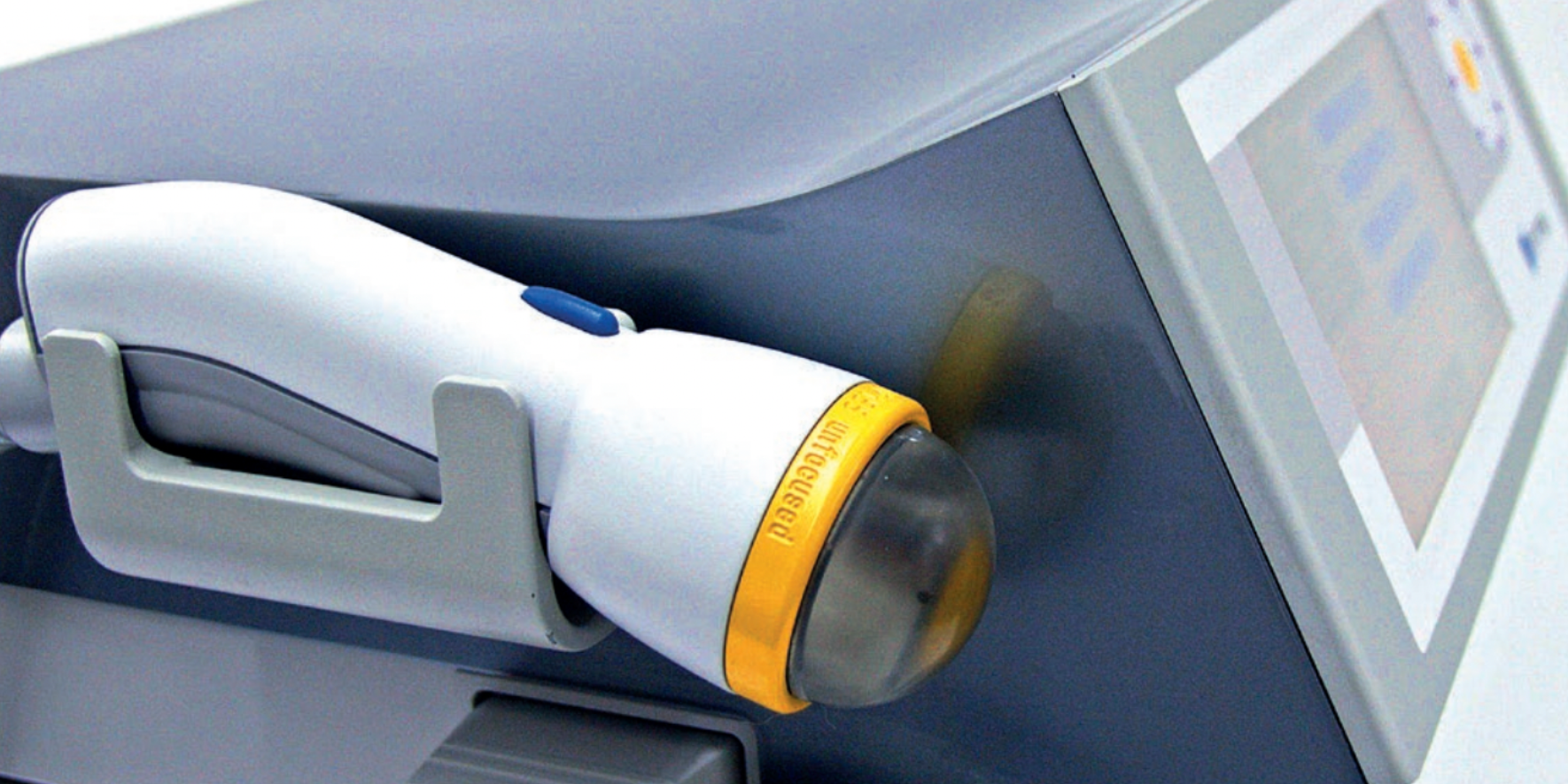

Pelvic Function Level 2C - Satellite - Long Beach CA - October 24 - 25 2026
Oct 24 2026 - Oct 25 2026




Pediatrics Level 1 - Treatment of Bowel and Bladder Disorders - Remote Course - October 24 - 25 2026
Oct 24 2026 - Oct 25 2026

Oncology of the Pelvic Floor Level 2B - Remote Course - October 24 - 25 2026
Oct 24 2026 - Oct 25 2026


Pelvic Function Level 2B - In-Person - Cape Cod MA - October 31 - November 1 2026
Oct 31 2026 - Nov 1 2026

Pelvic Function Level 1 - Satellite - St Augustine FL - October 31 - November 1 2026
Oct 31 2026 - Nov 1 2026



Menopause Transitions and Pelvic Rehab - Remote Course - November 7 - 8 2026
Nov 7 2026 - Nov 8 2026


Pelvic Function Level 1 - In-Person - Nashville TN - November 7 - 8 2026 - SOLD OUT
Nov 7 2026 - Nov 8 2026



Pharmacologic Considerations for the Pelvic Health Provider - Remote Course - November 7 2026
Nov 7 2026
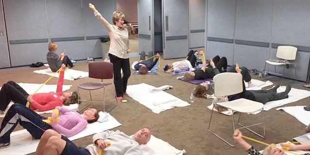

Rehabilitative Ultrasound Imaging Pelvic Health Satellite Lab Course - Self-Hosted - November 13 - 15 2026
Nov 13 2026 - Nov 15 2026