Pelvic Rehab Report
The official Herman & Wallace blog. New blogs post every Friday on topics relating to the field of pelvic floor dysfunction.

Herman & Wallace currently has satellite courses, remote courses, and online courses offered through our partner, Medbridge. These online courses provide education and patient engagement tools for pelvic floor dysfunction. H&W faculty have put together a collection of online continuing education courses with our partners at MedBridge. These convenient learning resources and can be purchased individually or as part of an annual subscription
The truth is that we all have hectic busy schedules that can make setting aside time for a live course can be difficult. Annual subscribers get access to all 800+ courses on the Medbridge site, their Home Exercise Program, and Patient Engagement platforms! As a Herman & Wallace referral, you are eligible for a discounted subscription with access to all of the content at MedBridge with promo code HWoverview.
Heather Rader, PT, DPT, PRPC, BCB-PMD recorded a new series for Medbridge last summer. Her courses explore:
- anatomical relevance of the pelvic girdle and pelvic floor structures
- common movement dysfunctions in rehabilitation
- overview of the sacroiliac joint, pubic symphysis, and coccyx
- bladder, bowel, and sexual dysfunctions created by pelvic floor dysfunctions
- connections between orthopedic dysfunction seen with pelvic floor dysfunctions
- orthopedic conditions such as back pain, hip pain, orthopedic trauma, and surgery
- diastasis recti abdominis cluster with pelvic floor dysfunctions, such as incontinence and pelvic pain syndromes in clinical practice
- including several other topics
The Medbridge online course catalog contains an in-depth physical therapy section with many familiar instructors including Holly Tanner and Steven Dischiavi. These online courses are a great way for pelvic rehabilitation specialists to gain knowledge and skills and complete CEU requirements without having to commit to a predetermined schedule.
Herman & Wallace continuing education courses are developed and instructed by our nationally recognized faculty members. Contact hours vary by course and may apply towards continuing education requirements according to each state.

The following is our interview with Jazma Dobbins PT, DPT, PRPC, CAPP-Pelvic. Jazma recently passed the Pelvic Rehabilitation Practitioner Certification (PRPC) exam. She practices in at TherapySouth in Gadsen, AL and is a Teaching Assistant for local Alabama satellite courses with H&W. Jazma was kind enough to share some thoughts about her career with us. Thank you, Jazma - and congratulations on receiving your PRPC!
Q: How did you get involved in the pelvic rehabilitation field?
A: Educating others about ways to empower themselves through healthcare advocacy is extremely important to me. I knew I wanted to be a pelvic health physical therapist the moment I read a similarly titled article in a women’s health magazine over 10 years ago. I was experiencing painful intercourse and urge incontinence. I was 20 years old and felt ashamed, abnormal, and alone. I had been given the unfortunate advice of “try drinking wine to relax” and “never drink alcohol if you have urge incontinence." So many inconsistencies to my young and frustrated mind.
That is when it dawned on me that I could do that, I could be a pelvic health physical therapist. Then we would have that service in my community. I was certain, and rightly so, that there were plenty of other women (I did not yet know of men’s health issues) who needed these services.
Q: What has your educational journey as a pelvic rehab therapist looked like?
A: I started my pelvic rehab education journey as a third-year student in PT school. I took the pelvic health level 1 course through the then called Section on Women’s Health. Needless to say, I was hooked on pelvic rehab. Over the following year, I completed the APTA pelvic series and earned my CAPP-Pelvic. In 2019 I did the Coccydynia and painful sitting course through Herman and Wallace and fell in love with H&W! Since then, I have had the pleasure of being a Teaching Assistant for multiple H&W courses.
Q: What do you love about assisting at courses?
A: What I love most about serving as a teaching assistant is the opportunity to learn from the participants. I have yet to TA a course where I didn’t learn a new skill or thought process. I love to TA the pelvic level 1 course most of all because of the eagerness and anticipation around learning a whole new world of what PT can do for our clients. I love to spread energy and passion and help create an environment of truly nonjudgmental learning.
Q: What is your message to course participants who are just starting their journey?
A: Keep pursuing your passion! Let no learning opportunity go unutilized and never stop learning!

This blog contains excerpts from an interview with Tara Sullivan, PT, DPT, PRPC, WCS, IF. Tara started in the healthcare field as a massage therapist, practicing over ten years including three years of teaching massage and anatomy and physiology. Tara has specialized exclusively in Pelvic Floor Dysfunction treating bowel, bladder, sexual dysfunctions, and pelvic pain since 2012.
Acute pain can indicate specific injury to the body. Chronic pain is very different. With Chronic Pelvic Pain (CPP) the initial injury has healed, but the pain continues because of changes in the nervous system, muscles, and tissues. Recognizing that the nervous system influences pain perception, especially in the chronic pelvic pain population, is the first step in treating these patients, but is it enough? Tara Sullivan and Alyson Lowrey are presenting a new remote course on chronic pelvic pain called Pain Science for the Chronic Pelvic Pain Population scheduled for July 17-18, 2021.
The medical definition of pain is an unpleasant feeling that is conveyed to the brain by sensory neurons. Pain is a universal experience that serves to alert the brain to potential damage to the body. It performs the function of triggering avoidance to preserve itself from harm. Oddly, the strength and unpleasantness of pain is not directly related to the nature or extent of the damage.
When the pain signal remains active in the nervous system for longer than six months and persists after the triggering event has healed, then it is cataloged as chronic pain. There is another layer when experiencing chronic pain known as central sensitization. This is an increased responsiveness of the nervous system that results in hypersensitivity and an increased pain response outside the area of injury. Pain itself can produce systematic and chemical brain changes resulting in more pain from fewer stimuli.
The course, Pain Science for the Chronic Pelvic Pain Population, offers tools to recognize when sensitization may be playing a role and provides the framework needed to apply pain science to the chronic pelvic pain population. In this course, you will gain an understanding and expand your knowledge on how pain science specifically presents in patients suffering from endometriosis, interstitial cystitis, primary dysmenorrhea, pelvic floor muscle overactivity, vulvodynia/vestibulodynia, vaginismus, and prostatitis.
Case studies and specific intervention techniques, including how to explain pain to a patient, are discussed so participants leave with the confidence to address the missing link in treating your patient’s chronic pelvic pain. We will also discuss how common rehab interventions such as manual therapy, dry needling, biofeedback, graded exposure, and therapeutic exercise assist in downregulating the nervous system.
On July 17-18th, 2021, Alyson Lowrey and Tara Sullivan team up to give you their combined experience of orthopedic and pelvic health in treating this population in the course Pain Science for the Chronic Pelvic Pain Population.

This blog contains excerpts from an interview with Pamela A. Downey, PT, DPT, WCS, BCB-PMD, PRPC, Pamela is a Board Certified Clinical Specialist in Women’s Health Physical Therapy and Board Certified in Biofeedback for Pelvic Muscle Dysfunction. She is the owner of Partnership in Therapy, private practice in Coral Gables, Florida. Dr. Downey's treatment focuses are pelvic floor dysfunction, urogynecological and colorectal issues, spine dysfunction, osteoporosis, and complaints associated with pregnancy and postpartum. Her mission is to educate and integrate healthy lifestyles for patients on the road to wellness.
Physical therapists often require special training to treat pudendal neuralgia. Pamela A. Downey is partnering with H&W to teach the Pudendal Neuralgia and Nerve Entrapment Remote Course, scheduled for June 19-20, 2021. This course teaches pudendal neuralgia diagnostic skills for practitioners to have an improved impact in treating patients with pudendal nerve/pelvic floor muscle dysfunctions.
Pudendal neuralgia is also known as Alcock’s syndrome, pudendal canal syndrome, or cyclist syndrome. This condition is caused by tension, compression, or entrapment of the pudendal nerve, and leads to pelvic pain, sexual dysfunction, difficulty with urination and defecation, among other issues.
Pudendal neuralgia is often unrecognized by physicians, including gynecologists, urologists, and neurologists. Dr. Downey observes that “Organizing your clinical decision-making process is key in determining the source of seated pain. Pudendal neuralgia can be a chicken and egg clinical phenomenon. My success comes from relying on a solid anatomy background in helping solve the pudendal puzzle.”
Successful treatments can include connective tissue mobilization, neural mobilization, and a home exercise program. Poor movement patterns can contribute to the symptoms of pudendal neuralgia. Physical therapy evaluation in these cases can include movement assessment and a gentle internal assessment of the patient's pelvic muscles. This provides information about the muscles’ ability to contract and relax. Exercises recommended to relax the pudendal nerve and provide temporary relief include cobra pose, side-lying hip abduction and extension, and wide-leg bridges.
Dr. Downey shares that she loves teaching the Pudendal Neuralgia and Nerve Entrapment Remote Course. “We teach the participants, in real-time, how to use evidence-based criteria to see if pudendal neuralgia makes sense as the driving diagnosis. Then we develop this confidence by careful dissection of case studies of real patients treated out over multiple visits, just like you do in the clinic."
Hone your decision-making process and gain confidence in the Pudendal Neuralgia and Nerve Entrapment Remote Course to treat pelvic pain with Pamela Downey on June 19-20, 2021.

Working with Physiatry for Pelvic Pain is a new remote course created by Dr. Allyson Shrikhande, scheduled for Jun 27, 2021. This course overviews the synergistic nature of pelvic physiatry with pelvic floor physical therapy, in hopes of promoting collaboration for the care of male and female chronic pelvic pain patients.
Dr. Allyson Shrikhande is a board-certified Physical Medicine and Rehabilitation specialist and is the Chair of the Medical Education Committee for the International Pelvic Pain Society. Allyson has published peer-reviewed articles on the treatment of muscle pain in academic journals and works closely with renowned pelvic pain gynecologists and urologists. Taking a team approach, she works with specialists in pelvic floor physical therapy, kinetics and movement, as well as acupuncturists, nutritionists, cognitive-behavioral therapists, and functional medicine physicians.
The following is our interview with Allyson Shrikhande on physiatry.
Q: What is a physiatrist?
A: A physiatrist is an MD or DO with a specialty in Physical Medicine and Rehabilitation. This non-operative medical discipline involves focusing on the neuromusculoskeletal system to help patients recover their functional well-being and quality of life. We describe physiatry as an extension of physical therapy because a physiatrist diagnoses, manages, and treats pain from injury, illness, or medical conditions, incorporating other methods in concert with physical therapy to rehabilitate the body. Physiatrists are trained not solely in one organ system – rather, they take a holistic, full-body approach that accounts for the interplay of different organ systems, both with each other and with the neuromuscular and myofascial systems.
Q: What does a physiatrist do?
A: Physiatrists work with physical therapy to rehabilitate the neuromuscular system. A core underlying theme in physiatry is the concept of Neuroplasticity. This is the understanding that the nervous system has the ability to form and reorganize synaptic connections, especially in response to experience or learning following injury.
Q: What do physiatrists treat?
A: Because physiatrists focus on the interconnected systems of the entire body, they treat a wide range of injuries and disorders. Physiatrists commonly work with patients who have pelvic, back or neck pain who are recovering from issues such as sports injuries, neuromuscular disorders, arthritis, or injuries to the brain or spinal cord.
Q: Why would I see a physiatrist?
A: At Pelvic Rehabilitation Medicine, our Pelvic Physiatrists diagnose and treat the structures of the pelvis – the muscles, nerves, and joints. One of our physiatrists can provide non-operative options to medically manage and treat pelvic pain and pelvic floor muscle dysfunction. We treat an array of symptoms under the umbrella of pelvic pain which includes pain with intercourse, urinary urgency/frequency or pain with urination, constipation or painful bowel movements, and pain affecting the coccyx, groin, pelvis, lower back, and lower abdomen.
Q: As a pelvic floor physical therapist, what can I learn from a physiatrist?
A: The relationship between physiatry and physical therapy is vital to the collaborative approach that our pelvic pain patients require. Physiatrists perform a full neuromuscular exam (including an internal pelvic floor exam) and can order imaging, prescribe oral medications, suppositories, and topical medications for some patients. Our physiatrists can also perform safe outpatient ultrasound-guided procedures to treat underlying neuromuscular dysfunction, all in combination with continued pelvic floor PT when appropriate.

Megan Pribyl, PT, CMPT is a practicing physical therapist at the Olathe Medical Center in Olathe, KS treating a diverse outpatient population in orthopedics including pelvic rehabilitation. Megan’s longstanding passion for both nutritional sciences and manual therapy has culminated in the creation of her remote course, Nutrition Perspectives for the Pelvic Rehab Therapist, designed to propel understanding of human physiology as it relates to pelvic conditions, pain, healing, and therapeutic response. She harnesses her passion to continually update this course with cutting-edge discoveries creating a unique experience sure to elevate your level of appreciation for the complex and fascinating nature of clinical presentations in orthopedic manual therapy and pelvic rehabilitation.
As a course developer and instructor for the Herman & Wallace Pelvic Rehab Institute, it is a privilege to continue sharing my passion for nutrition and pelvic rehabilitation with professionals nationwide. Interest in the topic continues to grow, and many pelvic rehab providers have identified nutrition as the “missing link” in their clinical practice. Nutrition Perspectives for the Pelvic Rehab Therapist has helped hundreds of pelvic rehab professionals integrate nutrition-related information into their clinical practice since 2015.

In the realm of nutrition, few questions provoke discussion with the same fervor as our title question: Organic Food vs. Conventional: Is There Any Difference? This question deserves a multi-dimensional answer - not unlike many topics in nutrition - including accessibility concerns, ethical factors for farmers, socio-economic factors, and our unique agricultural construct here in the United States. But the question about organic vs. conventional might just be the most important one deserving a thoughtful discussion to unravel the complexities around the topic of food.
You see, the answer to this question has profound implications for us. As we expand our ability to identify potential root contributors to conditions commonly encountered in pelvic rehabilitation, we must factor in nutrition. At first glance, it might be a stretch to see how one might link organic foods and potential effects on conditions such as constipation, inflammatory bowel diseases, IBS, PBS, and endometriosis for example. However, looking at food in a functional way, we acknowledge there may be under-appreciated qualitative differences between foods grown organically or produced conventionally.
Take, for example, the recent article by Kesse-Guyot et.al., 2020. which discusses the prospective association between organic food consumption and the risk of type 2 diabetes. In this study of over 30,000 participants, those with the highest quintile of organic food consumption compared to those with the lowest quintile had a 35% lower risk of having type 2 diabetes. The conclusion made by the authors was that organic food consumption was inversely associated with the risk of type 2 diabetes.
Said a different way, the study described a phenomenon where, for example, you might eat an organic bowl of oatmeal for breakfast and I might eat the same serving size conventional bowl of oatmeal for breakfast. If we extrapolate the comparison over our entire dietary intake pattern, you would have a 35% lower risk for developing type 2 diabetes compared to me…..despite you and I “eating the same foods”. How can this be possible? And might this begin to explain the sheer exasperation and frustration that can evolve in persons trying to make positive dietary changes - only to find they have no notable effect? How many times do you hear someone say “I am trying to eat healthily but it doesn’t seem to make a difference”.

Keeping in the context of type 2 diabetes, it is very well established that reductions in the richness and diversity of healthy microbes inhabiting the large intestine (gut dysbiosis) are correlative to metabolic syndrome. In those with type 2 diabetes, microbiomes showed a decrease in anti-inflammatory, probiotic, and other [beneficial] bacteria that could be pathogenic. (Das et al, 2021) Appreciating the differences between organic vs conventional - it is also well established that organic foods do carry less residue of herbicides and pesticides. These residues - which are found in higher concentration in conventionally produced foods - have been implicated in the same reduction in richness and diversity of microorganisms in the gut - which is contributory to dysbiosis. (Rueda-Ruzafa et all, 2019) Therefore it now seems not just plausible - but probable that there is a distinguishable difference between organic and conventional diets - to a degree at which all health care providers would do well to take notice.
In a report on the history of organic agriculture, author George Kuepper points out that:
“Pioneers of the organic movement believed that healthy food produced healthy people and that healthy people were the basis for a healthy society.”
And if organic foods can be a part of that, our patients deserve to know that these scientifically documented differences exist.
As our awareness of the connection between nutrition and health grows, so does the need to follow the science to share evidence-based and evidence-informed information. It is now more important than ever to have a working knowledge of nutrition basics as a pelvic rehabilitation professional. Plan to join us at one of our upcoming remote offerings of “Nutrition Perspectives for the Pelvic Rehab Therapist”: June 19-20 where we will explore this and many additional - and fascinating facets of the nutrition discussion.
Das, T., Jayasudha, R., Chakravarthy, S., Prashanthi, G. S., Bhargava, A., Tyagi, M., . . . Shivaji, S. (2021). Alterations in the gut bacterial microbiome in people with type 2 diabetes mellitus and diabetic retinopathy. Sci Rep, 11(1), 2738. doi:10.1038/s41598-021-82538-0
Kesse-Guyot, E., Rebouillat, P., Payrastre, L., Alles, B., Fezeu, L. K., Druesne-Pecollo, N., . . . Baudry, J. (2020). Prospective association between organic food consumption and the risk of type 2 diabetes: findings from the NutriNet-Sante cohort study. Int J Behav Nutr Phys Act, 17(1), 136. doi:10.1186/s12966-020-01038-y
Kuepper, George. (2010) A Brief Overview of the History and Philosophy of Organic Agriculture. Kerr Center for Sustainable Agriculture. http://kerrcenter.com/wp-content/uploads/2014/08/organic-philosophy-report.pdf Accessed May 14, 2021.
Rueda-Ruzafa, L., Cruz, F., Roman, P., & Cardona, D. (2019). Gut microbiota and neurological effects of glyphosate. Neurotoxicology, 75, 1-8. doi:10.1016/j.neuro.2019.08.006
Images:Par, Cecilia for Unsplash.
USDA organic seal.svg. Public Domain.

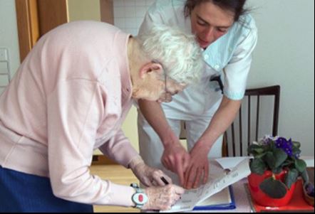
Deb Gulbrandson, PT, DPT has been a physical therapist for over 42 years with experience in acute care, home health, pediatrics, geriatrics, sports medicine, and consulting to business and industry. Dr. Gulbrandson frequently presents community talks on topics related to Osteoporosis and safe ways to develop Core Strength. She is a member of the APTA Geriatric and Private Practice Sections, a Certified Osteoporosis Exercise Specialist using the Meeks Method, and is a CEEAA (Certified Exercise Expert for the Aging Adult) through the Geriatric Section of the APTA.
Hello, my name is Deb Gulbrandson. May is National Osteoporosis Month, and my colleague, Frank Ciuba, and I are creators of the upcoming remote course Osteoporosis Management on June 12-13, 2021.
Did you know that approximately half of all women and a quarter of all men will break a bone due to osteoporosis? Equally disheartening, every year about one-third of adults in the US age 65 and older will fall. Many of these falls will result in broken bones.
Frank and I developed the Osteoporosis Management course to bring treatment protocols to practitioners in an organized and easily implementable format. This course is based on the Meeks Method created by Sara Meeks, PT, MS, GCS, with whom Frank and I taught for several years. With Sara’s blessing, we have branched out to add information on sleep hygiene, exercise dosing, and basic nutrition for a person with low bone mass. Knowing how to recognize signs, screen for osteoporosis, and design an effective and safe program can be life-changing for these patients.
In all likelihood, you are working with osteoporosis patients right now whether you know it or not. Osteoporosis affects all ages and stages of life including young and middle-aged men and women. Often we see patients with orthopedic or neurologic diagnoses who also have osteoporosis- known or unknown. Being able to address patients’ comorbidities helps ensure safety and quality of life. Many of the techniques we use can apply across several patient demographics. After all, who doesn’t benefit from postural alignment, back extensor/glute strengthening, and balance activities?
Working with this population has been extremely rewarding, because similar to pelvic health patients, these patients have been told there’s nothing you can do about it. They are incredibly grateful to find out that there IS something you can do about osteoporosis. They enthusiastically take an active role in their rehabilitation.
In addition, as a private practice owner for over 30 years, I’ve found osteoporosis to be a great business opportunity. An incredible niche practice, bringing in patients who have actively sought out my clinic with their self-referrals or by word of mouth.
We hope that you will join us this June 12-13 for our course Osteoporosis Management for an opportunity to experience posture, power, visual cueing, strengthening, and balance activities along with osteoporosis screening, evaluation, and evidence-based practice.
In this blog, instructor Heather Rader, PT, DPT, PRPC, BCB-PMD, discusses the concerns and fears therapists have when treating mothers recovering from childbirth and how her upcoming course can prepare therapists to confidently treat patients within hours to weeks after childbirth.
When is it OK for a new mom to start therapy?
When a new pelvic therapist asks me this question at courses, my answer begins by shifting the focus towards the birth itself. Childbirth is a mechanism of injury, not a “special population.” The real question then is when is it OK to start therapy after perineal or abdominal tissue trauma? When your clinical reasoning begins from this point of view, it removes biases you may have about childbirth and instills confidence in your skills as a trained clinician. Every therapist learns acute care skills - how to treat wounds, how to mobilize post-op patients, and how to screen for medical complications. Simply put, dear clinician, you know more than you think you do.
On average, 3.75 million women give birth in the US per year (1.) 100% will have some level of injury because of it. Those injuries will be perineal, abdominal, or both. There will be muscle strains and ligamentous sprains. There will be soft tissue bruising and swelling.
There are risks of immobility and re-injury without proper patient education. There might be stitches or staples in the abdominal or vulvar skin. There will be pain issues, mobility issues, safety issues, and definitely body mechanics and ergonomic issues. Given these obvious musculoskeletal and mobility impairments, I ask you to ponder this - what profession is better prepared to assess the acute and sub-acute needs of a new mother than rehab professionals?
Let’s imagine the same question posed to an acute care therapist about newly injured trauma patients.
Every lecture on the history of rehabilitation highlights the early days when bed rest was thought to be therapeutic.
Through research and clinical observations, we know immobility was, in some cases, deadly. Nowadays, it is considered standard of care to begin mobilization as early as medically possible, even in the ICU. And so should it be with new mothers. When should a person who was in an accident start rehab? What information would our therapist need to determine when to start and what early intervention is medically appropriate?
- Is the patient medically stable?
- Are the injured tissues structurally stable?
- Have any problems developed as a result of being immobilized in the hospital?
- Are there other co-morbidities to complicate the recovery?
- How is the patient’s mental status after the trauma?
- What social support is in place to assist with short-term and long-term needs?
- Can the patient function independently? Need home health? Benefit from outpatient therapy?
The clinical decision-making and critical thinking necessary to manage the care of an acute care patient is not much different than that of a pelvic therapist managing the recovery of a new or “acute” mother. More and more hospitals and birthing centers are incorporating acute care therapy within hours of birth. There are anatomical and physiological differences because of the effects of pregnancy itself that the clinician must learn, however. While the patient is recovering from childbirth, the body is returning to its pre-pregnancy state. Having a better understanding of the late pregnancy, birth, and the peripartum state has on healing can assist the motivated clinician in adding maternal-based therapy to their skill set.
The course Peripartum Advanced Topics covers medical screening, early exercise, patient education, hospital-based programming, and treatment strategies, as well as early outpatient care and fitness transition planning, such as returning to running.
If you are contemplating expanding your outpatient practice to see patients early in the “4th Trimester” or even earlier in the acute setting after the 4th stage of labor (recovery), consider signing up for the course.
- 1. https://www.cdc.gov/nchs/fastats/births.htm accessed 5/19/2021 10:30 AM

Mia Fine, MS, LMFT, CST, CIIP has written and instructing her remote course, Sexual Interviewing for Pelvic Health Therapists, with H&W on June 5-6, 2021. Mia (they/she) is a student of Queer Theory, Intersectionality, and Social Justice, and offers holistic, anti-oppressive, and trauma-informed therapy. This is a course intended for pelvic rehab therapists who want to learn tools and strategies from a sex therapist’s toolkit who work with patients experiencing pelvic pain, pelvic floor hypertonicity, and other pelvic floor concerns.
Let’s talk terms: Dyspareunia and Vaginismus
- Vaginismus: involuntary contraction of vaginal muscles preventing insertion/penetration
- Dyspareunia: pain felt during penetration
Symptoms (not limited to): pain - painful penetration, painful orgasms, painful periods, painful pelvic exams, inability to use tampons/cups, urinary or bowel hesitancy, feeling “too tight”
Causes (not limited to): stress, relationship concerns, mood concerns (anxiety, depression), insufficient arousal/desire/interest, trauma, side effects of meds, negative attitudes towards sex, and other pelvic pain concerns such as a tilted uterus.
The deal is, when it comes to dyspareunia and vaginismus there’s a cycle that can be difficult to break. The cycle is: you feel pain, then you feel broken (shame about feeling pain), then anxiety that pain will happen again, then you tighten your pelvic floor, and the cycle repeats. It becomes a self-fulfilling prophecy.
Often unwanted sexual pain goes unaddressed. Why? Because we are not taught about the interactions between feelings, relationships, and our body. We are not taught that sex should not be painful; that pain is (likely) our body giving us information that something is going on (Hello crappy sex education and the stigma of sexual health and body awareness!). It’s not uncommon that most people who experience sexual pain often feel they are broken.
You are not broken
How to heal from unwanted sexual pain? There’s a trifecta! Effective healing comes from working with a sex-positive medical provider, sex therapist, and pelvic floor PT. We will all collaborate!
Sex is not supposed to be painful. You are not broken.
Mia ️
#therapy #sextherapy #wellness #health #awareness #mentalhealth #bodyawareness
Steve Dischiavi holds credentials as a licensed physical therapist and a certified athletic trainer. He also has a manual therapy certification from the Ola Grimsby Institute and is board certified by the American Physical Therapy Association as a Sports Clinical Specialist (SCS).
The blog you’re about to read is targeted at clinicians just like myself. I was taught in PT school to evaluate the sacroiliac joint (SIJ) via a movement-based analysis. I even went on for a certification in manual therapy nearly 2 decades ago, and again was taught the movement-based approach. Well, as the song goes… “times they are a changin”….
So… if you’re a clinician who will diagnosis the SIJ as being “unstable” from movement-based testing, using tests that evaluate the positional movement of the PSIS… then you need to begin to question the approach you’re taking.
There is a contemporary conversation taking place within the field of physical therapy. If you’re like me and learned to evaluate the sacroiliac joint (SIJ) through movement-based testing, you need to be involved in this evolving conversation.
A great place to start is with a “Perspective” article published in Physical Therapy in 2019. It is titled “Changing the Narrative in Diagnosis and Management of Pain in the Sacroiliac Joint Area” written by Thorvaldur Palsson, et.al.1
The narrative dives into several areas pertaining to the treatment of this mysterious region of the body. It gives the reader a thoughtful and contemplative view of the contemporary conversation surrounding the evaluation and treatment of the SIJ.
The target audience should be the clinicians who continue to evaluate the SIJ solely on movement based analysis, attempting to diagnosis the SIJ with terms related to movement dysfunction. We have all heard terms like “upslip, downslip, or sacral torsions”. The narrative addresses why this is not the best approach to diagnosing the SIJ.
The article also touches on the concepts of “pain science” and how assessing how the SIJ “moves” tells the clinician very little about why the tissues about the SIJ might be sensitized. The article scratches the surface about the patient’s pain experience and how harmful terms such as “instability” and how the SIJ “goes out” are ways that can perpetuate and even heighten the pain experience for your patients.
The Perspective article gives the reader a great introduction as to why you might consider altering your approach to the SIJ. I’ll leave you with a quote directly from the article:
“If clinical decisions are based on a construct that lacks plausibility and clinical tests lacking in validity and reliability, the entire management paradigm must be questioned.”
The next question you might be asking yourself… ok, how do I learn a new management paradigm? A great start would be to take the 4-hour introductory webinar, Sacroiliac Joint Current Concepts - Remote Course - June 26, 2021, offered by Herman and Wallace… learn the evidence-based approach to the SIJ with Steve Dischiavi, PT, DPT, MPT, SCS, ATC, COMT who has been working closely on the hip and pelvis for over 20 years!
I hope to see you at the webinar!
1. Palsson TS, Gibson W, Darlow B, et al. Changing the Narrative in Diagnosis and Management of Pain in the Sacroiliac Joint Area. Phys Ther. 2019;99(11):1511-1519.
By accepting you will be accessing a service provided by a third-party external to https://hermanwallace.com/
All Upcoming Continuing Education Courses


Pelvic Function Level 1 - Satellite - Des Moines IA - July 26 - 27 2025 - SOLD OUT
Jul 26 2025 - Jul 27 2025

Pelvic Function Level 1 - Satellite - Minneapolis MN - July 26 - 27 2025 - SOLD OUT
Jul 26 2025 - Jul 27 2025

Pelvic Function Level 1 - Satellite - Paso Robles CA - July 26 - 27 2025 - SOLD OUT
Jul 26 2025 - Jul 27 2025

Pelvic Function Level 1 - Satellite - St. Augustine FL - July 26 - 27 2025
Jul 26 2025 - Jul 27 2025

Pelvic Function Level 1 - Satellite - Virginia Beach VA - July 26 - 27 2025 - SOLD OUT
Jul 26 2025 - Jul 27 2025


Pelvic Function Level 1 - In-Person - Chicago IL - August 2 - 3 2025 - SOLD OUT
Aug 2 2025 - Aug 3 2025


Menopause Transitions and Pelvic Rehab - Remote Course - August 9 - 10 2025
Aug 9 2025 - Aug 10 2025


Pelvic Function Level 2C - Satellite - Palm Beach FL - August 16 - 17 2025
Aug 16 2025 - Aug 17 2025

Pelvic Function Level 2C - Satellite - New Orleans LA - August 16 - 17 2025
Aug 16 2025 - Aug 17 2025
Pelvic Function Level 2C - Satellite - Paso Robles CA - August 16 - 17 2025
Aug 16 2025 - Aug 17 2025


Pediatrics Level 1 - Treatment of Bowel and Bladder Disorders - Remote Course - August 23 - 24 2025
Aug 23 2025 - Aug 24 2025

Pelvic Function Level 1 - Satellite - Jacksonville FL - August 23 - 24 2025
Aug 23 2025 - Aug 24 2025

Pelvic Function Level 1 - Satellite - Chicago IL - August 23 - 24 2025 - SOLD OUT
Aug 23 2025 - Aug 24 2025


Pelvic Function Level 1 - Satellite - Hermosa Beach CA - August 23 - 24 2025
Aug 23 2025 - Aug 24 2025

Rehabilitative Ultrasound Imaging Pelvic Health Satellite Lab Course - Self-Hosted - September 5 - 7 2025
Sep 5 2025 - Sep 7 2025
Rehabilitative Ultrasound: Orthopedic Topics Satellite Lab Course - Self-Hosted - September 5 - 6 2025
Sep 5 2025 - Sep 6 2025

Rehabilitative Ultrasound Imaging Pelvic Health Satellite Lab Course - Indianapolis IN - September 5 - 7 2025
Sep 5 2025 - Sep 7 2025
Rehabilitative Ultrasound: Orthopedic Topics Satellite Lab Course - Indianapolis IN - September 5 - 6 2025
Sep 5 2025 - Sep 6 2025

Rehabilitative Ultrasound Imaging: Women's Health - Satellite Lab Course - Seattle WA - September 5 - 7 2025
Sep 5 2025 - Sep 7 2025
Rehabilitative Ultrasound Imaging: Orthopedic Topics Satellite Lab Course - Seattle WA - September 5 - 6 2025
Sep 5 2025 - Sep 6 2025

Pelvic Function Level 1 - Satellite - Sacramento CA - September 6 - 7 2025 - SOLD OUT
Sep 6 2025 - Sep 7 2025


Pelvic Function Level 1 - Satellite - San Francisco CA - September 6 - 7 2025 - SOLD OUT
Sep 6 2025 - Sep 7 2025